The Solution Configurations of Inactive and Activated Dntr Have Implications for the Sliding Dimer Mechanism of Lysr Transcription Factors.
Lerche, M., Dian, C., Round, A., Lonneborg, R., Brzezinski, P., Leonard, G.A.(2016) Sci Rep 6: 19988
- PubMed: 26817994 Search on PubMedSearch on PubMed Central
- DOI: https://doi.org/10.1038/srep19988
- Primary Citation Related Structures:
5AE4, 5AE5 - PubMed Abstract:
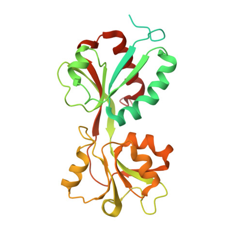
LysR Type Transcriptional Regulators (LTTRs) regulate basic metabolic pathways or virulence gene expression in prokaryotes. Evidence suggests that the activation of LTTRs involves a conformational change from an inactive compact apo- configuration that represses transcription to an active, expanded holo- form that promotes it. However, no LTTR has yet been observed to adopt both configurations. Here, we report the results of structural studies of various forms of the LTTR DntR. Crystal structures of apo-DntR and of a partially autoinducing mutant H169T-DntR suggest that active and inactive DntR maintain a compact homotetrameric configuration. However, Small Angle X-ray Scattering (SAXS) studies on solutions of apo-, H169T- and inducer-bound holo-DntR indicate a different behaviour, suggesting that while apo-DntR maintains a compact configuration in solution both H169T- and holo-DntR adopt an expanded conformation. Models of the SAXS-obtained solution conformations of apo- and holo-DntR homotetramers in complex with promoter-operator region DNA are consistent with previous observations of a shifting of LTTR DNA binding sites upon activation and a consequent relaxation in the bend of the promoter-operator region DNA. Our results thus provide clear evidence at the molecular level which strongly supports the 'sliding dimer' hypothesis concerning LTTR activation mechanisms.
- Structural Bioloy Group, European Synchrotron Radiation Facility (ESRF), CS 40220, 38043 Grenoble Cedex 9, France.
Organizational Affiliation: