5ADP | pdb_00005adp

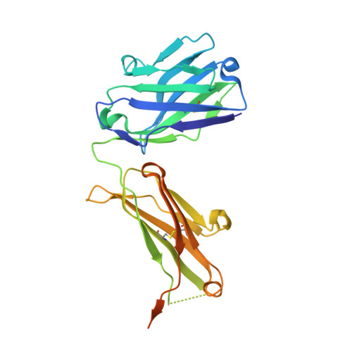
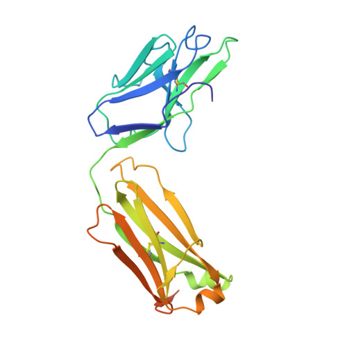
Crystal structure of the A.17 antibody FAB fragment - Light chain S35R mutant


Experimental Data Snapshot

  • Method: X-RAY DIFFRACTION
  • Resolution: 2.13 Å
  • R-Value Free: 
    0.220 (Depositor), 0.221 (DCC) 
  • R-Value Work: 
    0.179 (Depositor), 0.182 (DCC) 
  • R-Value Observed: 
    0.181 (Depositor) 

Starting Model: experimental
View more details

wwPDB Validation 3D Report Full Report

Validation slider image for 5ADP

This is version 1.3 of the entry. See complete history. 

Literature

Robotic Qm/Mm-Driven Maturation of Antibody Combining Sites.

Smirnov, I.V., Golovin, A.V., Chatziefthimiou, S.D., Stepanova, A.V., Peng, Y., Zolotareva, O.I., Belogurov, A.A., Kurkova, I.N., Ponomarenko, N.A., Wilmanns, M., Blackburn, G.M., Gabibov, A.G., Lerner, R.A.

(2016) Sci Adv 2: 01695

  • DOI: https://doi.org/10.1126/sciadv.1501695
  • Primary Citation Related Structures: 
    5ADO, 5ADP

  • PubMed Abstract: 

    In vitro selection of antibodies from large repertoires of immunoglobulin (Ig) combining sites using combinatorial libraries is a powerful tool, with great potential for generating in vivo scavengers for toxins. However, addition of a maturation function is necessary to enable these selected antibodies to more closely mimic the full mammalian immune response. We approached this goal using quantum mechanics/molecular mechanics (QM/MM) calculations to achieve maturation in silico. We preselected A17, an Ig template, from a naïve library for its ability to disarm a toxic pesticide related to organophosphorus nerve agents. Virtual screening of 167,538 robotically generated mutants identified an optimum single point mutation, which experimentally boosted wild-type Ig scavenger performance by 170-fold. We validated the QM/MM predictions via kinetic analysis and crystal structures of mutant apo-A17 and covalently modified Ig, thereby identifying the displacement of one water molecule by an arginine as delivering this catalysis.


  • Organizational Affiliation: 
    • Shemyakin-Ovchinnikov Institute of Bioorganic Chemistry, Russian Academy of Sciences, ulitsa Miklukho-Maklaya 16/10, 117997 Moscow V-437, Russian Federation.

Macromolecule Content 

  • Total Structure Weight: 54.08 kDa 
  • Atom Count: 3,467 
  • Modeled Residue Count: 428 
  • Deposited Residue Count: 502 
  • Unique protein chains: 2

Macromolecules

Find similar proteins by:|  3D Structure
Entity ID: 1
MoleculeChains  Sequence LengthOrganismDetailsImage
FAB A.17A [auth H]255Homo sapiensMutation(s): 0 
Entity Groups
Sequence Clusters30% Identity50% Identity70% Identity90% Identity95% Identity100% Identity
Sequence Annotations
Expand
Reference Sequence
Find similar proteins by:|  3D Structure
Entity ID: 2
MoleculeChains  Sequence LengthOrganismDetailsImage
FAB A.17B [auth L]247Homo sapiensMutation(s): 0 
Entity Groups
Sequence Clusters30% Identity50% Identity70% Identity90% Identity95% Identity100% Identity
Sequence Annotations
Expand
Reference Sequence

Experimental Data & Validation

Experimental Data

  • Method: X-RAY DIFFRACTION
  • Resolution: 2.13 Å
  • R-Value Free:  0.220 (Depositor), 0.221 (DCC) 
  • R-Value Work:  0.179 (Depositor), 0.182 (DCC) 
  • R-Value Observed: 0.181 (Depositor) 
Space Group: P 1 21 1
Unit Cell:
Length ( Å )Angle ( ˚ )
a = 52.547α = 90
b = 67.04β = 107.63
c = 66.95γ = 90
Software Package:
Software NamePurpose
PHENIXrefinement
XDSdata reduction
Aimlessdata scaling
MOLREPphasing

Structure Validation

View Full Validation Report


Entry History 

Revision History  (Full details and data files)

  • Version 1.0: 2016-11-09
    Type: Initial release
  • Version 1.1: 2019-07-17
    Changes: Data collection
  • Version 1.2: 2024-01-10
    Changes: Data collection, Database references, Other, Refinement description
  • Version 1.3: 2024-11-20
    Changes: Structure summary