5A80 | pdb_00005a80

Crystal structure of human JMJD2A in complex with compound 40


Experimental Data Snapshot

  • Method: X-RAY DIFFRACTION
  • Resolution: 2.28 Å
  • R-Value Free: 
    0.249 (Depositor), 0.253 (DCC) 
  • R-Value Work: 
    0.190 (Depositor), 0.197 (DCC) 
  • R-Value Observed: 
    0.193 (Depositor) 

Starting Model: experimental
View more details

wwPDB Validation 3D Report Full Report

Validation slider image for 5A80

Ligand Structure Quality Assessment 


This is version 1.3 of the entry. See complete history

Literature

Docking and Linking of Fragments to Discover Jumonji Histone Demethylase Inhibitors.

Korczynska, M.Le, D.D.Younger, N.Gregori-Puigjane, E.Tumber, A.Krojer, T.Velupillai, S.Gileadi, C.Nowak, R.P.Iwasa, E.Pollock, S.B.Ortiz Torres, I.Oppermann, U.Shoichet, B.K.Fujimori, D.G.

(2016) J Med Chem 59: 1580

  • DOI: https://doi.org/10.1021/acs.jmedchem.5b01527
  • Primary Citation Related Structures: 
    5A7N, 5A7O, 5A7P, 5A7Q, 5A7S, 5A7W, 5A80

  • PubMed Abstract: 

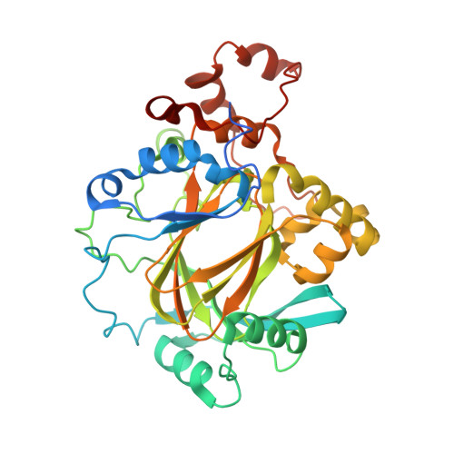
    Development of tool molecules that inhibit Jumonji demethylases allows for the investigation of cancer-associated transcription. While scaffolds such as 2,4-pyridinedicarboxylic acid (2,4-PDCA) are potent inhibitors, they exhibit limited selectivity. To discover new inhibitors for the KDM4 demethylases, enzymes overexpressed in several cancers, we docked a library of 600,000 fragments into the high-resolution structure of KDM4A. Among the most interesting chemotypes were the 5-aminosalicylates, which docked in two distinct but overlapping orientations. Docking poses informed the design of covalently linked fragment compounds, which were further derivatized. This combined approach improved affinity by ∼ 3 log-orders to yield compound 35 (Ki = 43 nM). Several hybrid inhibitors were selective for KDM4C over the related enzymes FIH, KDM2A, and KDM6B while lacking selectivity against the KDM3 and KDM5 subfamilies. Cocrystal structures corroborated the docking predictions. This study extends the use of structure-based docking from fragment discovery to fragment linking optimization, yielding novel KDM4 inhibitors.


  • Organizational Affiliation
    • Structural Genomics Consortium, University of Oxford , Oxford OX3 7DQ, U.K.

Macromolecule Content 

  • Total Structure Weight: 90.46 kDa 
  • Atom Count: 5,779 
  • Modeled Residue Count: 696 
  • Deposited Residue Count: 762 
  • Unique protein chains: 1

Macromolecules

Find similar proteins by:|  3D Structure
Entity ID: 1
MoleculeChains  Sequence LengthOrganismDetailsImage
LYSINE-SPECIFIC DEMETHYLASE 4A
A, B
381Homo sapiensMutation(s): 0 
EC: 1.14.11 (PDB Primary Data), 1.14.11.66 (UniProt), 1.14.11.69 (UniProt)
UniProt & NIH Common Fund Data Resources
Find proteins for O75164 (Homo sapiens)
Explore O75164 
Go to UniProtKB:  O75164
PHAROS:  O75164
GTEx:  ENSG00000066135 
Entity Groups
Sequence Clusters30% Identity50% Identity70% Identity90% Identity95% Identity100% Identity
UniProt GroupO75164
Sequence Annotations
Expand
Reference Sequence

Small Molecules

Ligands 5 Unique
IDChains Name / Formula / InChI Key2D Diagram3D Interactions
9CJ

Query on 9CJ


Download:Ideal Coordinates CCD File
E [auth A],
N [auth B]
2-[5-[2-(3-methoxyphenyl)ethanoylamino]-2-oxidanyl-phenyl]pyridine-4-carboxylic acid
C21 H18 N2 O5
QSWJSOQQNWMPME-UHFFFAOYSA-N
SO4

Query on SO4


Download:Ideal Coordinates CCD File
C [auth A],
L [auth B]
SULFATE ION
O4 S
QAOWNCQODCNURD-UHFFFAOYSA-L
ZN

Query on ZN


Download:Ideal Coordinates CCD File
F [auth A],
O [auth B]
ZINC ION
Zn
PTFCDOFLOPIGGS-UHFFFAOYSA-N
EDO

Query on EDO


Download:Ideal Coordinates CCD File
G [auth A]
H [auth A]
I [auth A]
J [auth A]
K [auth A]
G [auth A],
H [auth A],
I [auth A],
J [auth A],
K [auth A],
P [auth B],
Q [auth B],
R [auth B],
S [auth B],
T [auth B]
1,2-ETHANEDIOL
C2 H6 O2
LYCAIKOWRPUZTN-UHFFFAOYSA-N
MN

Query on MN


Download:Ideal Coordinates CCD File
D [auth A],
M [auth B]
MANGANESE (II) ION
Mn
WAEMQWOKJMHJLA-UHFFFAOYSA-N

Experimental Data & Validation

Experimental Data

  • Method: X-RAY DIFFRACTION
  • Resolution: 2.28 Å
  • R-Value Free:  0.249 (Depositor), 0.253 (DCC) 
  • R-Value Work:  0.190 (Depositor), 0.197 (DCC) 
  • R-Value Observed: 0.193 (Depositor) 
Space Group: P 21 21 2
Unit Cell:
Length ( Å )Angle ( ˚ )
a = 101.81α = 90
b = 149.19β = 90
c = 57.24γ = 90
Software Package:
Software NamePurpose
PHENIXrefinement
XDSdata reduction
SCALAdata scaling
PHENIXphasing

Structure Validation

View Full Validation Report


Ligand Structure Quality Assessment 


Entry History 

Revision History  (Full details and data files)

  • Version 1.0: 2016-01-13
    Type: Initial release
  • Version 1.1: 2016-03-09
    Changes: Database references
  • Version 1.2: 2018-01-24
    Changes: Structure summary
  • Version 1.3: 2024-01-10
    Changes: Data collection, Database references, Derived calculations, Other, Refinement description