5A43 | pdb_00005a43

Crystal structure of a dual topology fluoride ion channel.


Experimental Data Snapshot

  • Method: X-RAY DIFFRACTION
  • Resolution: 2.58 Å
  • R-Value Free: 
    0.264 (Depositor), 0.268 (DCC) 
  • R-Value Work: 
    0.224 (Depositor), 0.238 (DCC) 
  • R-Value Observed: 
    0.226 (Depositor) 

wwPDB Validation 3D Report Full Report

Validation slider image for 5A43

Ligand Structure Quality Assessment 


This is version 1.3 of the entry. See complete history

Literature

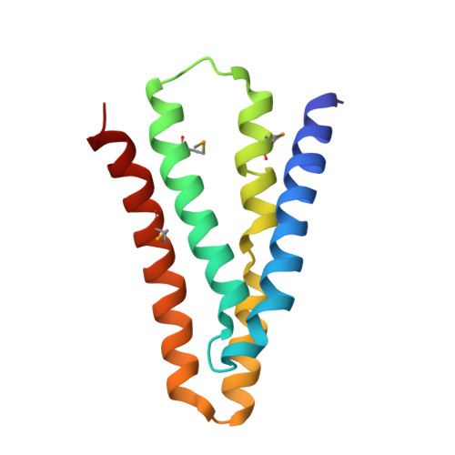
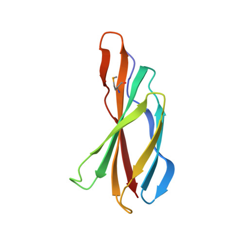
Crystal Structures of a Double-Barrelled Fluoride Ion Channel.

Stockbridge, R.B.Kolmakova-Partensky, L.Shane, T.Koide, A.Koide, S.Miller, C.Newstead, S.

(2015) Nature 525: 548

  • DOI: https://doi.org/10.1038/nature14981
  • Primary Citation Related Structures: 
    5A40, 5A43, 5NKQ

  • PubMed Abstract: 

    To contend with hazards posed by environmental fluoride, microorganisms export this anion through F(-)-specific ion channels of the Fluc family. Since the recent discovery of Fluc channels, numerous idiosyncratic features of these proteins have been unearthed, including strong selectivity for F(-) over Cl(-) and dual-topology dimeric assembly. To understand the chemical basis for F(-) permeation and how the antiparallel subunits convene to form a F(-)-selective pore, here we solve the crystal structures of two bacterial Fluc homologues in complex with three different monobody inhibitors, with and without F(-) present, to a maximum resolution of 2.1 Å. The structures reveal a surprising 'double-barrelled' channel architecture in which two F(-) ion pathways span the membrane, and the dual-topology arrangement includes a centrally coordinated cation, most likely Na(+). F(-) selectivity is proposed to arise from the very narrow pores and an unusual anion coordination that exploits the quadrupolar edges of conserved phenylalanine rings.


  • Organizational Affiliation
    • Department of Biochemistry, Howard Hughes Medical Institute, Brandeis University, Waltham, Massachusetts 02454, USA.

Macromolecule Content 

  • Total Structure Weight: 49.97 kDa 
  • Atom Count: 3,481 
  • Modeled Residue Count: 442 
  • Deposited Residue Count: 444 
  • Unique protein chains: 2

Macromolecules

Find similar proteins by:|  3D Structure
Entity ID: 1
MoleculeChains  Sequence LengthOrganismDetailsImage
PUTATIVE FLUORIDE ION TRANSPORTER CRCB
A, B
126Escherichia coli S88Mutation(s): 0 
Membrane Entity: Yes 
UniProt
Find proteins for B7LI20 (Escherichia coli O45:K1 (strain S88 / ExPEC))
Explore B7LI20 
Go to UniProtKB:  B7LI20
Entity Groups
Sequence Clusters30% Identity50% Identity70% Identity90% Identity95% Identity100% Identity
UniProt GroupB7LI20
Sequence Annotations
Expand
Reference Sequence
Find similar proteins by:|  3D Structure
Entity ID: 2
MoleculeChains  Sequence LengthOrganismDetailsImage
MONOBODIES
C, D
96Homo sapiensMutation(s): 0 

Experimental Data & Validation

Experimental Data

  • Method: X-RAY DIFFRACTION
  • Resolution: 2.58 Å
  • R-Value Free:  0.264 (Depositor), 0.268 (DCC) 
  • R-Value Work:  0.224 (Depositor), 0.238 (DCC) 
  • R-Value Observed: 0.226 (Depositor) 
Space Group: P 41
Unit Cell:
Length ( Å )Angle ( ˚ )
a = 87.42α = 90
b = 87.42β = 90
c = 146.81γ = 90
Software Package:
Software NamePurpose
PHENIXrefinement
MOSFLMdata reduction
SCALAdata scaling
SHELXphasing

Structure Validation

View Full Validation Report


Ligand Structure Quality Assessment 


Entry History 

Deposition Data

Revision History  (Full details and data files)

  • Version 1.0: 2015-09-02
    Type: Initial release
  • Version 1.1: 2015-09-23
    Changes: Database references
  • Version 1.2: 2015-09-30
    Changes: Database references
  • Version 1.3: 2024-11-13
    Changes: Data collection, Database references, Derived calculations, Other, Structure summary