5A37 | pdb_00005a37

Mutations in the Calponin homology domain of Alpha-Actinin-2 affect Actin binding and incorporation in muscle.


Experimental Data Snapshot

  • Method: X-RAY DIFFRACTION
  • Resolution: 1.88 Å
  • R-Value Free: 
    0.216 (Depositor), 0.220 (DCC) 
  • R-Value Work: 
    0.178 (Depositor), 0.187 (DCC) 
  • R-Value Observed: 
    0.180 (Depositor) 

wwPDB Validation 3D Report Full Report

Validation slider image for 5A37

This is version 1.2 of the entry. See complete history

Literature

Hypertrophic Cardiomyopathy Mutations in the Calponin-Homology Domain of Actn2 Affect Actin Binding and Cardiomyocyte Z-Disc Incorporation.

Haywood, N.Wolny, M.Rogers, B.Trinh, C.H.Shuping, Y.Edwards, T.A.Peckham, M.

(2016) Biochem J 473: 2485

  • DOI: https://doi.org/10.1042/BCJ20160421
  • Primary Citation Related Structures: 
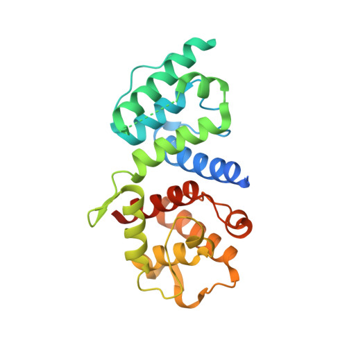
    5A36, 5A37, 5A38, 5A4B

  • PubMed Abstract: 

    α-Actinin-2 (ACTN2) is the only muscle isoform of α-actinin expressed in cardiac muscle. Mutations in this protein have been implicated in mild to moderate forms of hypertrophic cardiomyopathy (HCM). We have investigated the effects of two mutations identified from HCM patients, A119T and G111V, on the secondary and tertiary structure of a purified actin binding domain (ABD) of ACTN2 by circular dichroism and X-ray crystallography, and show small but distinct changes for both mutations. We also find that both mutants have reduced F-actin binding affinity, although the differences are not significant. The full length mEos2 tagged protein expressed in adult cardiomyocytes shows that both mutations additionally affect Z-disc localization and dynamic behaviour. Overall, these two mutations have small effects on structure, function and behaviour, which may contribute to a mild phenotype for this disease.


  • Organizational Affiliation
    • Astbury Centre for Structural Molecular Biology and the School of Molecular and Cellular Biology, Faculty of Biological Sciences, University of Leeds, Leeds LS2 9JT, U.K.

Macromolecule Content 

  • Total Structure Weight: 57.26 kDa 
  • Atom Count: 3,854 
  • Modeled Residue Count: 439 
  • Deposited Residue Count: 500 
  • Unique protein chains: 1

Macromolecules

Find similar proteins by:|  3D Structure
Entity ID: 1
MoleculeChains  Sequence LengthOrganismDetailsImage
HUMAN ALPHA-ACTININ-2
A, B
250Homo sapiensMutation(s): 1 
UniProt & NIH Common Fund Data Resources
Find proteins for P35609 (Homo sapiens)
Explore P35609 
Go to UniProtKB:  P35609
PHAROS:  P35609
GTEx:  ENSG00000077522 
Entity Groups
Sequence Clusters30% Identity50% Identity70% Identity90% Identity95% Identity100% Identity
UniProt GroupP35609
Sequence Annotations
Expand
Reference Sequence

Experimental Data & Validation

Experimental Data

  • Method: X-RAY DIFFRACTION
  • Resolution: 1.88 Å
  • R-Value Free:  0.216 (Depositor), 0.220 (DCC) 
  • R-Value Work:  0.178 (Depositor), 0.187 (DCC) 
  • R-Value Observed: 0.180 (Depositor) 
Space Group: P 1
Unit Cell:
Length ( Å )Angle ( ˚ )
a = 38.24α = 73.68
b = 46.49β = 80.08
c = 70.38γ = 75.53
Software Package:
Software NamePurpose
REFMACrefinement
iMOSFLMdata reduction
SCALAdata scaling
REFMACphasing

Structure Validation

View Full Validation Report


Entry History 

Deposition Data

Revision History  (Full details and data files)

  • Version 1.0: 2016-06-22
    Type: Initial release
  • Version 1.1: 2016-08-24
    Changes: Database references
  • Version 1.2: 2024-05-08
    Changes: Data collection, Database references, Other