5A2E

Extracellular SRCR domains of human CD6


Experimental Data Snapshot

  • Method: X-RAY DIFFRACTION
  • Resolution: 3.15 Å
  • R-Value Free: 0.283 
  • R-Value Work: 0.253 
  • R-Value Observed: 0.255 

wwPDB Validation   3D Report Full Report


Ligand Structure Quality Assessment 


This is version 1.3 of the entry. See complete history


Literature

Structures of Cd6 and its Ligand Cd166 Give Insight Into Their Interaction.

Chappell, P.E.Garner, L.I.Yan, J.Metcalfe, C.Hatherley, D.Johnson, S.Robinson, C.V.Lea, S.M.Brown, M.H.

(2015) Structure 23: 1426

  • DOI: https://doi.org/10.1016/j.str.2015.05.019
  • Primary Citation of Related Structures:  
    5A2E, 5A2F

  • PubMed Abstract: 

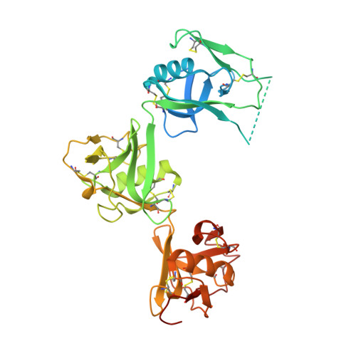
    CD6 is a transmembrane protein with an extracellular region containing three scavenger receptor cysteine rich (SRCR) domains. The membrane proximal domain of CD6 binds the N-terminal immunoglobulin superfamily (IgSF) domain of another cell surface receptor, CD166, which also engages in homophilic interactions. CD6 expression is mainly restricted to T cells, and the interaction between CD6 and CD166 regulates T-cell activation. We have solved the X-ray crystal structures of the three SRCR domains of CD6 and two N-terminal domains of CD166. This first structure of consecutive SRCR domains reveals a nonlinear organization. We characterized the binding sites on CD6 and CD166 and showed that a SNP in CD6 causes glycosylation that hinders the CD6/CD166 interaction. Native mass spectrometry analysis showed that there is competition between the heterophilic and homophilic interactions. These data give insight into how interactions of consecutive SRCR domains are perturbed by SNPs and potential therapeutic reagents.


  • Organizational Affiliation

    Sir William Dunn School of Pathology, University of Oxford, South Parks Road, Oxford OX1 3RE, UK.


Macromolecules
Find similar proteins by:  (by identity cutoff)  |  3D Structure
Entity ID: 1
MoleculeChains Sequence LengthOrganismDetailsImage
T-CELL DIFFERENTIATION ANTIGEN CD6371Homo sapiensMutation(s): 0 
UniProt & NIH Common Fund Data Resources
Find proteins for P30203 (Homo sapiens)
Explore P30203 
Go to UniProtKB:  P30203
PHAROS:  P30203
GTEx:  ENSG00000013725 
Entity Groups  
Sequence Clusters30% Identity50% Identity70% Identity90% Identity95% Identity100% Identity
UniProt GroupP30203
Sequence Annotations
Expand
  • Reference Sequence
Experimental Data & Validation

Experimental Data

  • Method: X-RAY DIFFRACTION
  • Resolution: 3.15 Å
  • R-Value Free: 0.283 
  • R-Value Work: 0.253 
  • R-Value Observed: 0.255 
  • Space Group: P 63 2 2
Unit Cell:
Length ( Å )Angle ( ˚ )
a = 161.48α = 90
b = 161.48β = 90
c = 93.85γ = 120
Software Package:
Software NamePurpose
BUSTERrefinement
XDSdata reduction
Aimlessdata scaling
PHASERphasing

Structure Validation

View Full Validation Report


Ligand Structure Quality Assessment 


Entry History 

Deposition Data

Revision History  (Full details and data files)

  • Version 1.0: 2015-07-22
    Type: Initial release
  • Version 1.1: 2015-08-19
    Changes: Database references
  • Version 1.2: 2020-07-29
    Type: Remediation
    Reason: Carbohydrate remediation
    Changes: Data collection, Derived calculations, Other, Structure summary
  • Version 1.3: 2024-01-10
    Changes: Data collection, Database references, Refinement description, Structure summary