4ZY6 | pdb_00004zy6

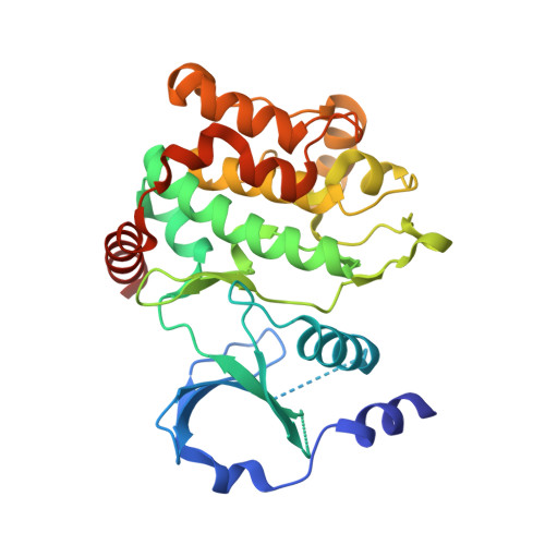
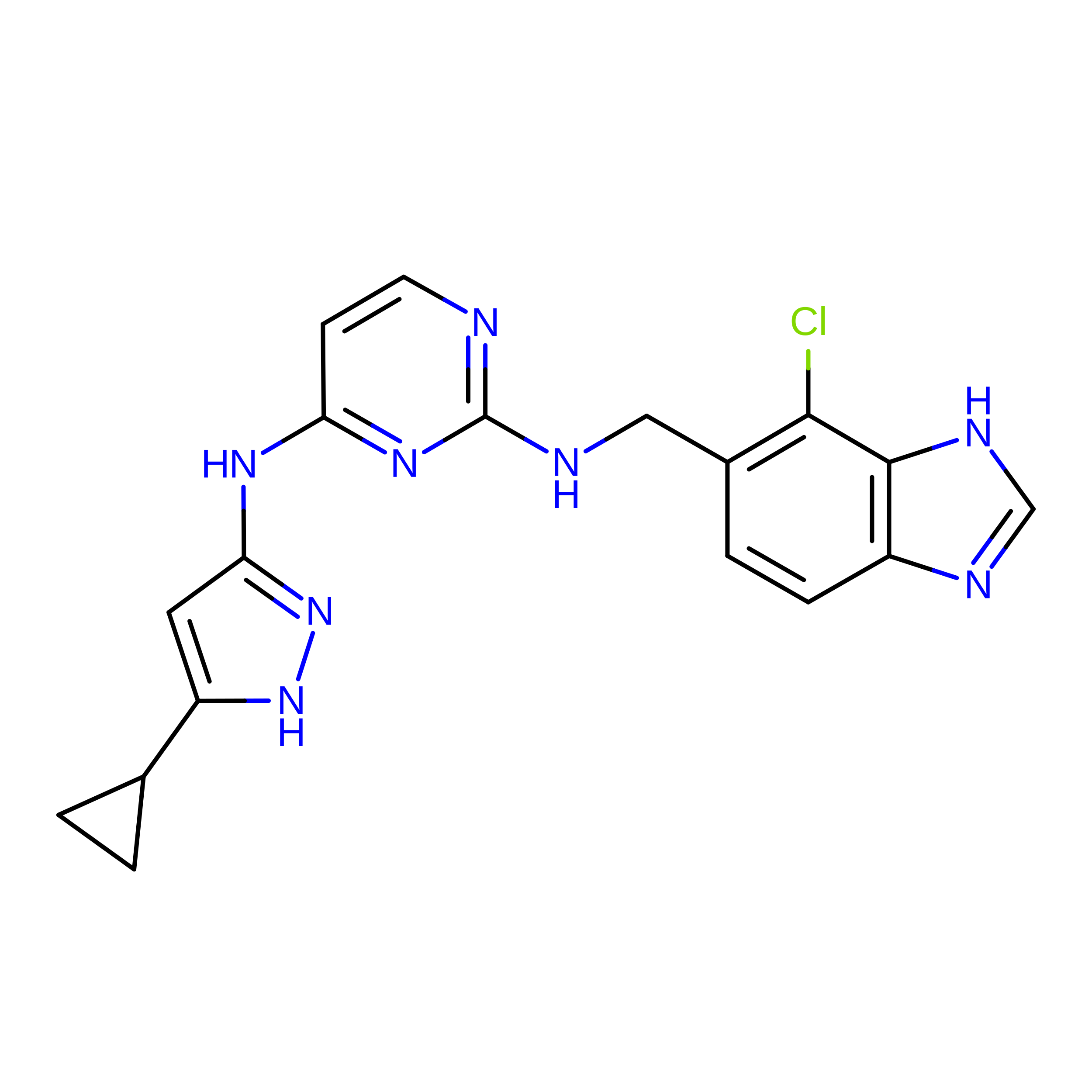
Crystal structure of P21-activated kinase 1 in complex with an inhibitor compound 29


Experimental Data Snapshot

  • Method: X-RAY DIFFRACTION
  • Resolution: 2.15 Å
  • R-Value Free: 
    0.239 (Depositor), 0.236 (DCC) 
  • R-Value Work: 
    0.187 (Depositor), 0.183 (DCC) 
  • R-Value Observed: 
    0.190 (Depositor) 

wwPDB Validation 3D Report Full Report

Validation slider image for 4ZY6

Ligand Structure Quality Assessment 


This is version 1.3 of the entry. See complete history. 

Literature

Structure-Guided Design of Group I Selective p21-Activated Kinase Inhibitors.

Crawford, J.J., Lee, W., Aliagas, I., Mathieu, S., Hoeflich, K.P., Zhou, W., Wang, W., Rouge, L., Murray, L., La, H., Liu, N., Fan, P.W., Cheong, J., Heise, C.E., Ramaswamy, S., Mintzer, R., Liu, Y., Chao, Q., Rudolph, J.

(2015) J Med Chem 58: 5121-5136

  • DOI: https://doi.org/10.1021/acs.jmedchem.5b00572
  • Primary Citation Related Structures: 
    4ZY4, 4ZY5, 4ZY6, 5BMS

  • PubMed Abstract: 

    The p21-activated kinases (PAKs) play important roles in cytoskeletal organization, cellular morphogenesis, and survival and have generated significant attention as potential therapeutic targets for cancer. Following a high-throughput screen, we identified an aminopyrazole scaffold-based series that was optimized to yield group I selective PAK inhibitors. A structure-based design effort aimed at targeting the ribose pocket for both potency and selectivity led to much-improved group I vs II selectivity. Early lead compounds contained a basic primary amine, which was found to be a major metabolic soft spot with in vivo clearance proceeding predominantly via N-acetylation. We succeeded in identifying replacements with improved metabolic stability, leading to compounds with lower in vivo rodent clearance and excellent group I PAK selectivity.


  • Organizational Affiliation: 
    • #Shanghai Chempartner Inc., 998 Halei Road, Zhangjiang Hi-Tech Park, Pudong New Area, Shanghai 201203, People's Republic of China.

Macromolecule Content 

  • Total Structure Weight: 68 kDa 
  • Atom Count: 4,707 
  • Modeled Residue Count: 564 
  • Deposited Residue Count: 602 
  • Unique protein chains: 1

Macromolecules

Find similar proteins by:|  3D Structure
Entity ID: 1
MoleculeChains  Sequence LengthOrganismDetailsImage
Serine/threonine-protein kinase PAK 1
A, B
301Homo sapiensMutation(s): 1 
Gene Names: PAK1
EC: 2.7.11.1
UniProt & NIH Common Fund Data Resources
Find proteins for Q13153 (Homo sapiens)
Explore Q13153 
Go to UniProtKB:  Q13153
PHAROS:  Q13153
GTEx:  ENSG00000149269 
Entity Groups
Sequence Clusters30% Identity50% Identity70% Identity90% Identity95% Identity100% Identity
UniProt GroupQ13153
Sequence Annotations
Expand
Reference Sequence

Experimental Data & Validation

Experimental Data

  • Method: X-RAY DIFFRACTION
  • Resolution: 2.15 Å
  • R-Value Free:  0.239 (Depositor), 0.236 (DCC) 
  • R-Value Work:  0.187 (Depositor), 0.183 (DCC) 
  • R-Value Observed: 0.190 (Depositor) 
Space Group: P 1 21 1
Unit Cell:
Length ( Å )Angle ( ˚ )
a = 62.634α = 90
b = 82.241β = 106.11
c = 66.137γ = 90
Software Package:
Software NamePurpose
BUSTERrefinement
HKL-2000data reduction
HKL-2000data scaling
PHASERphasing

Structure Validation

View Full Validation Report


Ligand Structure Quality Assessment 


Entry History 

Deposition Data

Revision History  (Full details and data files)

  • Version 1.0: 2015-07-01
    Type: Initial release
  • Version 1.1: 2015-07-15
    Changes: Database references
  • Version 1.2: 2015-12-16
    Changes: Refinement description
  • Version 1.3: 2024-11-13
    Changes: Data collection, Database references, Derived calculations, Structure summary