4ZTT

Crystal structures of ferritin mutants reveal diferric-peroxo intermediates


Experimental Data Snapshot

  • Method: X-RAY DIFFRACTION
  • Resolution: 1.83 Å
  • R-Value Free: 0.192 
  • R-Value Work: 0.156 
  • R-Value Observed: 0.158 

wwPDB Validation   3D Report Full Report


This is version 1.3 of the entry. See complete history


Literature

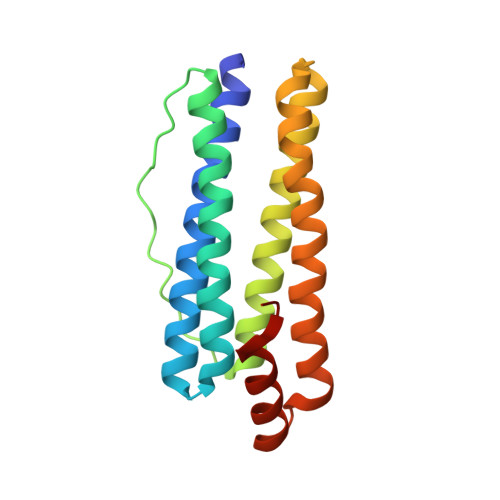
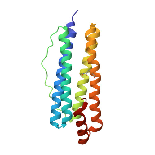
Structural Basis of Novel Iron-Uptake Route and Reaction Intermediates in Ferritins from Gram-Negative Bacteria.

Kim, S.Lee, J.H.Seok, J.H.Park, Y.H.Jung, S.W.Cho, A.E.Lee, C.Chung, M.S.Kim, K.H.

(2016) J Mol Biol 428: 5007-5018

  • DOI: https://doi.org/10.1016/j.jmb.2016.10.022
  • Primary Citation of Related Structures:  
    4XGS, 4ZTT, 5C6F

  • PubMed Abstract: 

    Iron and oxygen chemistry is mediated by iron proteins for many biological functions. Carboxylate-bridged diiron enzymes including ferritin have the common mechanism of oxygen activation via peroxodiferric intermediates. However, the route for iron uptake and the structural identification of intermediates still remain incomplete. The 4-fold symmetry channel of Helicobacter pylori ferritin was previously proposed as the iron-uptake route in eubacteria, but the amino acid residues at the 4-fold channel are not highly conserved. Here, we show evidence for a short path for iron uptake from His93 on the surface to the ferroxidase center in H. pylori ferritin and Escherichia coli ferritin. The amino acid residues along this path are highly conserved in Gram-negative bacteria and some archaea, and the mutants containing S20A and H93L showed significantly decreased iron oxidation. Surprisingly, the E. coli ferritin S20A crystal structure showed oxygen binding and side-on, symmetric μ-η 2 2 peroxodiferric and oxodiferric intermediates. The results provide the structural basis for understanding the chemical nature of intermediates in iron oxidation in bacteria and some of archaea.


  • Organizational Affiliation

    Department of Biotechnology & Bioinformatics, Korea University, Sejong 339-700, Korea.


Macromolecules
Find similar proteins by:  (by identity cutoff)  |  3D Structure
Entity ID: 1
MoleculeChains Sequence LengthOrganismDetailsImage
Bacterial non-heme ferritin
A, B, C, D, E
A, B, C, D, E, F
166Escherichia coli DH1Mutation(s): 1 
Gene Names: ftnAZ2960ECs2613
EC: 1.16.3.2
UniProt
Find proteins for P0A998 (Escherichia coli (strain K12))
Explore P0A998 
Go to UniProtKB:  P0A998
Entity Groups  
Sequence Clusters30% Identity50% Identity70% Identity90% Identity95% Identity100% Identity
UniProt GroupP0A998
Sequence Annotations
Expand
  • Reference Sequence
Small Molecules
Ligands 8 Unique
IDChains Name / Formula / InChI Key2D Diagram3D Interactions
FEO
Query on FEO

Download Ideal Coordinates CCD File 
FA [auth E],
G [auth A],
L [auth B]
MU-OXO-DIIRON
Fe2 O
NPMYUMBHPJGBFA-UHFFFAOYSA-N
GOL
Query on GOL

Download Ideal Coordinates CCD File 
DA [auth D]
EA [auth D]
HA [auth E]
IA [auth E]
J [auth A]
DA [auth D],
EA [auth D],
HA [auth E],
IA [auth E],
J [auth A],
K [auth A],
O [auth B],
P [auth B],
QA [auth F],
U [auth C],
V [auth C],
W [auth C],
X [auth C],
Y [auth C]
GLYCEROL
C3 H8 O3
PEDCQBHIVMGVHV-UHFFFAOYSA-N
FE2
Query on FE2

Download Ideal Coordinates CCD File 
JA [auth E],
R [auth C]
FE (II) ION
Fe
CWYNVVGOOAEACU-UHFFFAOYSA-N
FE
Query on FE

Download Ideal Coordinates CCD File 
AA [auth D],
KA [auth F],
LA [auth F],
Q [auth C],
Z [auth D]
FE (III) ION
Fe
VTLYFUHAOXGGBS-UHFFFAOYSA-N
PER
Query on PER

Download Ideal Coordinates CCD File 
BA [auth D]PEROXIDE ION
O2
ANAIPYUSIMHBEL-UHFFFAOYSA-N
OXY
Query on OXY

Download Ideal Coordinates CCD File 
S [auth C]OXYGEN MOLECULE
O2
MYMOFIZGZYHOMD-UHFFFAOYSA-N
OH
Query on OH

Download Ideal Coordinates CCD File 
CA [auth D]
GA [auth E]
H [auth A]
I [auth A]
M [auth B]
CA [auth D],
GA [auth E],
H [auth A],
I [auth A],
M [auth B],
N [auth B],
OA [auth F],
PA [auth F],
T [auth C]
HYDROXIDE ION
H O
XLYOFNOQVPJJNP-UHFFFAOYSA-M
O
Query on O

Download Ideal Coordinates CCD File 
MA [auth F],
NA [auth F]
OXYGEN ATOM
O
XLYOFNOQVPJJNP-UHFFFAOYSA-N
Experimental Data & Validation

Experimental Data

  • Method: X-RAY DIFFRACTION
  • Resolution: 1.83 Å
  • R-Value Free: 0.192 
  • R-Value Work: 0.156 
  • R-Value Observed: 0.158 
  • Space Group: I 4
Unit Cell:
Length ( Å )Angle ( ˚ )
a = 128.816α = 90
b = 128.816β = 90
c = 172.247γ = 90
Software Package:
Software NamePurpose
REFMACrefinement
XDSdata processing
XDSdata scaling
PHENIXphasing

Structure Validation

View Full Validation Report


Entry History 

Deposition Data

Revision History  (Full details and data files)

  • Version 1.0: 2016-06-15
    Type: Initial release
  • Version 1.1: 2016-11-16
    Changes: Database references
  • Version 1.2: 2017-04-05
    Changes: Database references
  • Version 1.3: 2020-02-19
    Changes: Data collection