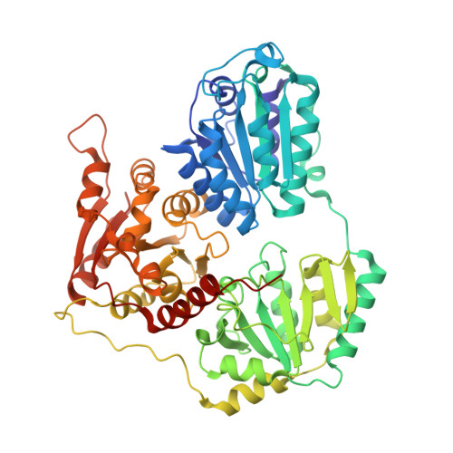
4ZP1

Crystal structure of Zymomonas mobilis pyruvate decarboxylase variant Glu473Ala


Experimental Data Snapshot

  • Method: X-RAY DIFFRACTION
  • Resolution: 2.21 Å
  • R-Value Free: 0.217 
  • R-Value Work: 0.170 
  • R-Value Observed: 0.172 

wwPDB Validation   3D Report Full Report


Ligand Structure Quality Assessment 


This is version 1.2 of the entry. See complete history


Literature

Tuning and Switching Enantioselectivity of Asymmetric Carboligation in an Enzyme through Mutational Analysis of a Single Hot Spot.

Wechsler, C.Meyer, D.Loschonsky, S.Funk, L.M.Neumann, P.Ficner, R.Brodhun, F.Muller, M.Tittmann, K.

(2015) Chembiochem 16: 2580-2584

  • DOI: https://doi.org/10.1002/cbic.201500529
  • Primary Citation of Related Structures:  
    4ZP1

  • PubMed Abstract: 

    Enantioselective bond making and breaking is a hallmark of enzyme action, yet switching the enantioselectivity of the reaction is a difficult undertaking, and typically requires extensive screening of mutant libraries and multiple mutations. Here, we demonstrate that mutational diversification of a single catalytic hot spot in the enzyme pyruvate decarboxylase gives access to both enantiomers of acyloins acetoin and phenylacetylcarbinol, important pharmaceutical precursors, in the case of acetoin even starting from the unselective wild-type protein. Protein crystallography was used to rationalize these findings and to propose a mechanistic model of how enantioselectivity is controlled. In a broader context, our studies highlight the efficiency of mechanism-inspired and structure-guided rational protein design for enhancing and switching enantioselectivity of enzymatic reactions, by systematically exploring the biocatalytic potential of a single hot spot.


  • Organizational Affiliation

    Abt. Molekulare Enzymologie, Georg-August-Universität Göttingen, Justus-von-Liebig-Weg 11, 37077, Göttingen, Germany.


Macromolecules
Find similar proteins by:  (by identity cutoff)  |  3D Structure
Entity ID: 1
MoleculeChains Sequence LengthOrganismDetailsImage
Pyruvate decarboxylase
A, B, C, D
568Zymomonas mobilisMutation(s): 1 
Gene Names: pdcZMO1360
EC: 4.1.1.1
UniProt
Find proteins for P06672 (Zymomonas mobilis subsp. mobilis (strain ATCC 31821 / ZM4 / CP4))
Explore P06672 
Go to UniProtKB:  P06672
Entity Groups  
Sequence Clusters30% Identity50% Identity70% Identity90% Identity95% Identity100% Identity
UniProt GroupP06672
Sequence Annotations
Expand
  • Reference Sequence
Small Molecules
Ligands 4 Unique
IDChains Name / Formula / InChI Key2D Diagram3D Interactions
TPP
Query on TPP

Download Ideal Coordinates CCD File 
G [auth A],
K [auth B],
N [auth C],
P [auth D]
THIAMINE DIPHOSPHATE
C12 H19 N4 O7 P2 S
AYEKOFBPNLCAJY-UHFFFAOYSA-O
GOL
Query on GOL

Download Ideal Coordinates CCD File 
J [auth B],
M [auth C]
GLYCEROL
C3 H8 O3
PEDCQBHIVMGVHV-UHFFFAOYSA-N
NI
Query on NI

Download Ideal Coordinates CCD File 
F [auth A],
I [auth B]
NICKEL (II) ION
Ni
VEQPNABPJHWNSG-UHFFFAOYSA-N
MG
Query on MG

Download Ideal Coordinates CCD File 
E [auth A],
H [auth B],
L [auth C],
O [auth D]
MAGNESIUM ION
Mg
JLVVSXFLKOJNIY-UHFFFAOYSA-N
Experimental Data & Validation

Experimental Data

  • Method: X-RAY DIFFRACTION
  • Resolution: 2.21 Å
  • R-Value Free: 0.217 
  • R-Value Work: 0.170 
  • R-Value Observed: 0.172 
  • Space Group: P 1 21 1
Unit Cell:
Length ( Å )Angle ( ˚ )
a = 70.29α = 90
b = 167.64β = 101.66
c = 110.82γ = 90
Software Package:
Software NamePurpose
PHENIXrefinement
XSCALEdata scaling
PDB_EXTRACTdata extraction
XDSdata reduction
PHASERphasing

Structure Validation

View Full Validation Report


Ligand Structure Quality Assessment 


Entry History & Funding Information

Deposition Data


Funding OrganizationLocationGrant Number
German Research FoundationGermanyFOR 1296/TP3

Revision History  (Full details and data files)

  • Version 1.0: 2015-11-04
    Type: Initial release
  • Version 1.1: 2016-01-27
    Changes: Database references
  • Version 1.2: 2017-09-06
    Changes: Author supporting evidence