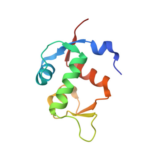
4ZFI

Structure of Mdm2 with low molecular weight inhibitor


Experimental Data Snapshot

  • Method: X-RAY DIFFRACTION
  • Resolution: 2.00 Å
  • R-Value Free: 0.243 
  • R-Value Work: 0.198 
  • R-Value Observed: 0.200 

wwPDB Validation   3D Report Full Report


Ligand Structure Quality Assessment 


This is version 1.1 of the entry. See complete history


Literature

A Unique Mdm2-Binding Mode of the 3-Pyrrolin-2-one- and 2-Furanone-Based Antagonists of the p53-Mdm2 Interaction.

Surmiak, E.Twarda-Clapa, A.Zak, K.M.Musielak, B.Tomala, M.D.Kubica, K.Grudnik, P.Madej, M.Jablonski, M.Potempa, J.Kalinowska-Tluscik, J.Domling, A.Dubin, G.Holak, T.A.

(2016) ACS Chem Biol 11: 3310-3318

  • DOI: https://doi.org/10.1021/acschembio.6b00596
  • Primary Citation of Related Structures:  
    4ZFI, 4ZGK

  • PubMed Abstract: 
  • The p53 pathway is inactivated in almost all types of cancer by mutations in the p53 encoding gene or overexpression of the p53 negative regulators, Mdm2 and/or Mdmx. Restoration of the p53 function by inhibition of the p53-Mdm2/Mdmx interaction opens up a prospect for a nongenotoxic anticancer therapy ...

    The p53 pathway is inactivated in almost all types of cancer by mutations in the p53 encoding gene or overexpression of the p53 negative regulators, Mdm2 and/or Mdmx. Restoration of the p53 function by inhibition of the p53-Mdm2/Mdmx interaction opens up a prospect for a nongenotoxic anticancer therapy. Here, we present the syntheses, activities, and crystal structures of two novel classes of Mdm2-p53 inhibitors that are based on the 3-pyrrolin-2-one and 2-furanone scaffolds. The structures of the complexes formed by these inhibitors and Mdm2 reveal the dimeric protein molecular organization that has not been observed in the small-molecule/Mdm2 complexes described until now. In particular, the 6-chloroindole group does not occupy the usual Trp-23 pocket of Mdm2 but instead is engaged in dimerization. This entirely unique binding mode of the compounds opens new possibilities for optimization of the Mdm2-p53 interaction inhibitors.


    Organizational Affiliation

    Malopolska Centre of Biotechnology, Jagiellonian University , Gronostajowa 7a, 30-387 Krakow, Poland.


Macromolecules
Find similar proteins by:  (by identity cutoff)  |  3D Structure
Entity ID: 1
MoleculeChainsSequence LengthOrganismDetailsImage
E3 ubiquitin-protein ligase Mdm2
A, B, C, D
97Homo sapiensMutation(s): 0 
Gene Names: MDM2
EC: 6.3.2 (PDB Primary Data), 2.3.2.27 (UniProt)
UniProt & NIH Common Fund Data Resources
Find proteins for Q00987 (Homo sapiens)
Explore Q00987 
Go to UniProtKB:  Q00987
PHAROS:  Q00987
Entity Groups  
Sequence Clusters30% Identity50% Identity70% Identity90% Identity95% Identity100% Identity
UniProt GroupQ00987
Protein Feature View
Expand
  • Reference Sequence
Small Molecules
Ligands 1 Unique
IDChainsName / Formula / InChI Key2D Diagram3D Interactions
4NJ
Query on 4NJ

Download Ideal Coordinates CCD File 
E [auth A],
F [auth B],
G [auth C],
H [auth D]
(5S)-3,5-bis(4-chlorobenzyl)-4-(6-chloro-1H-indol-3-yl)-5-hydroxy-1-methyl-1,5-dihydro-2H-pyrrol-2-one
C27 H21 Cl3 N2 O2
HHGSWONIEYVVCX-MHZLTWQESA-N
 Ligand Interaction
Binding Affinity Annotations 
IDSourceBinding Affinity
4NJ BindingDB:  4ZFI Kd: min: 500, max: 1000 (nM) from 2 assay(s)
Binding MOAD:  4ZFI Kd: 500 (nM) from 1 assay(s)
Experimental Data & Validation

Experimental Data

  • Method: X-RAY DIFFRACTION
  • Resolution: 2.00 Å
  • R-Value Free: 0.243 
  • R-Value Work: 0.198 
  • R-Value Observed: 0.200 
  • Space Group: P 21 21 21
Unit Cell:
Length ( Å )Angle ( ˚ )
a = 66.36α = 90
b = 70.25β = 90
c = 96.44γ = 90
Software Package:
Software NamePurpose
MOSFLMdata reduction
SCALAdata scaling
PHASERphasing
REFMACrefinement
PDB_EXTRACTdata extraction

Structure Validation

View Full Validation Report


Ligand Structure Quality Assessment 


Entry History & Funding Information

Deposition Data


Funding OrganizationLocationGrant Number
the Foundation for Polish SciencePolandTEAM Programme
the polish National Centre of SciencePolandUMO-2011/01/D/NZ1/01169

Revision History  (Full details and data files)

  • Version 1.0: 2016-10-19
    Type: Initial release
  • Version 1.1: 2016-12-28
    Changes: Database references