


Structure of Gan1D, a 6-phospho-beta-galactosidase from Geobacillus stearothermophilus, in complex with 6-phospho-glucose
Lansky, S., Zehavi, A., Dvir, H., Shoham, Y., Shoham, G.To be published.
Experimental Data Snapshot
Starting Model: experimental
View more details
Entity ID: 1 | |||||
|---|---|---|---|---|---|
| Molecule | Chains | Sequence Length | Organism | Details | Image |
| Putative 6-phospho-beta-galactobiosidase | 485 | Geobacillus stearothermophilus | Mutation(s): 0 Gene Names: gan1D EC: 3.2.1.85 | 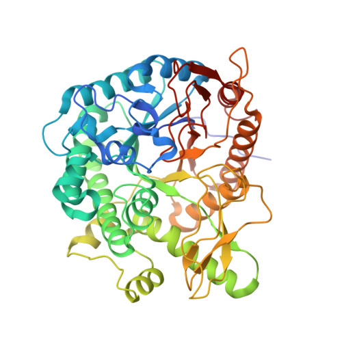 | |
UniProt | |||||
Entity Groups | |||||
| Sequence Clusters | 30% Identity50% Identity70% Identity90% Identity95% Identity100% Identity | ||||
| UniProt Group | W8QF82 | ||||
Sequence AnnotationsExpand | |||||
Reference Sequence | |||||
| Ligands 3 Unique | |||||
|---|---|---|---|---|---|
| ID | Chains | Name / Formula / InChI Key | 2D Diagram | 3D Interactions | |
| BG6 Download:Ideal Coordinates CCD File | C [auth A], G [auth B] | 6-O-phosphono-beta-D-glucopyranose C6 H13 O9 P NBSCHQHZLSJFNQ-VFUOTHLCSA-N |  | ||
| GOL Download:Ideal Coordinates CCD File | D [auth A], E [auth A], H [auth B], I [auth B] | GLYCEROL C3 H8 O3 PEDCQBHIVMGVHV-UHFFFAOYSA-N |  | ||
| IMD Download:Ideal Coordinates CCD File | F [auth A] | IMIDAZOLE C3 H5 N2 RAXXELZNTBOGNW-UHFFFAOYSA-O |  | ||
| Length ( Å ) | Angle ( ˚ ) |
|---|---|
| a = 107.158 | α = 90 |
| b = 68.944 | β = 100.39 |
| c = 152.896 | γ = 90 |
| Software Name | Purpose |
|---|---|
| REFMAC | refinement |
| HKL-3000 | data reduction |
| HKL-3000 | data scaling |
| PHASER | phasing |


