4Z9S | pdb_00004z9s

Non-covalent assembly of monoubiquitin that mimics K11 poly-ubiquitin


Experimental Data Snapshot

  • Method: X-RAY DIFFRACTION
  • Resolution: 2.30 Å
  • R-Value Free: 
    0.223 (Depositor), 0.238 (DCC) 
  • R-Value Work: 
    0.185 (Depositor), 0.193 (DCC) 

Starting Model: experimental
View more details

wwPDB Validation 3D Report Full Report

Validation slider image for 4Z9S

This is version 1.3 of the entry. See complete history

Literature

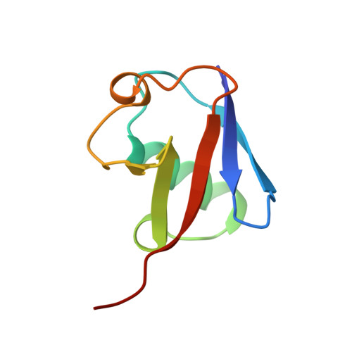
Tetrameric Assembly of Monoubiquitin Accurately Mimics the Lys11 Polyubiquitin Chain Structure.

Levin-Kravets, O.Shohat, N.Prag, G.

(2015) Biochemistry 54: 4704-4710

  • DOI: https://doi.org/10.1021/acs.biochem.5b00498
  • Primary Citation Related Structures: 
    4Z9S

  • PubMed Abstract: 

    Specific lysine residues on the ubiquitin surface were selected during the course of evolution to form different polyubiquitin chain structures that signal diverse cellular processes. A vast number of ubiquitin receptors specifically recognize and decode the signals conferred by these polyubiquitin chains. The mechanisms of formation and the structure of Lys11-linked ubiquitin, which signals for cell-cycle and innate immune control, have been elucidated. Here, we present a new crystal structure of monomeric ubiquitin that accurately mimics one of the structures of Lys11-linked ubiquitin. Analysis of the ubiquitin:ubiquitin interface demonstrates structural fitness and specificity. The interaction is exclusively hydrophilic, leaving the Ile44 hydrophobic patch, a major recognition site for ubiquitin receptors, exposed. These noncovalent ubiquitin:ubiquitin interactions are nearly identical to those reported for Lys11-linked ubiquitin and seem to play a significant role in stabilizing the crystal structure without the isopeptide bond. In vitro cross-linking analysis with wild-type ubiquitin or its mutants partially mimics the interactions in the crystal. We suggest that these interactions may play a biological role in transmitting Lys11-linked ubiquitin chain-type cellular signals.


  • Organizational Affiliation
    • Department of Biochemistry and Molecular Biology and Institute for Structural Biology, George S. Wise Faculty of Life Sciences, Tel Aviv University, Tel Aviv, Israel.

Macromolecule Content 

  • Total Structure Weight: 35.45 kDa 
  • Atom Count: 2,633 
  • Modeled Residue Count: 300 
  • Deposited Residue Count: 304 
  • Unique protein chains: 1

Macromolecules

Find similar proteins by:|  3D Structure
Entity ID: 1
MoleculeChains  Sequence LengthOrganismDetailsImage
Ubiquitin-40S ribosomal protein S27a
A, B, C, D
76Bos taurusMutation(s): 0 
Gene Names: UBC
UniProt
Find proteins for P62992 (Bos taurus)
Explore P62992 
Go to UniProtKB:  P62992
Entity Groups
Sequence Clusters30% Identity50% Identity70% Identity90% Identity95% Identity100% Identity
UniProt GroupP62992
Sequence Annotations
Expand
Reference Sequence

Small Molecules

Ligands 2 Unique
IDChains Name / Formula / InChI Key2D Diagram3D Interactions
MLA

Query on MLA


Download:Ideal Coordinates CCD File
E [auth A]
F [auth A]
K [auth B]
N [auth C]
O [auth C]
E [auth A],
F [auth A],
K [auth B],
N [auth C],
O [auth C],
R [auth D]
MALONIC ACID
C3 H4 O4
OFOBLEOULBTSOW-UHFFFAOYSA-N
SCN

Query on SCN


Download:Ideal Coordinates CCD File
G [auth A]
H [auth A]
I [auth A]
J [auth A]
L [auth B]
G [auth A],
H [auth A],
I [auth A],
J [auth A],
L [auth B],
M [auth B],
P [auth C],
Q [auth C],
S [auth D]
THIOCYANATE ION
C N S
ZMZDMBWJUHKJPS-UHFFFAOYSA-M

Experimental Data & Validation

Experimental Data

  • Method: X-RAY DIFFRACTION
  • Resolution: 2.30 Å
  • R-Value Free:  0.223 (Depositor), 0.238 (DCC) 
  • R-Value Work:  0.185 (Depositor), 0.193 (DCC) 
Space Group: C 1 2 1
Unit Cell:
Length ( Å )Angle ( ˚ )
a = 81.319α = 90
b = 81.301β = 129.33
c = 58.597γ = 90
Software Package:
Software NamePurpose
REFMACrefinement
MOSFLMdata reduction
SCALAdata scaling
MOLREPphasing

Structure Validation

View Full Validation Report


Entry History 

& Funding Information

Deposition Data


Funding OrganizationLocationGrant Number
Israeli Science Foundation464/11

Revision History  (Full details and data files)

  • Version 1.0: 2015-07-29
    Type: Initial release
  • Version 1.1: 2015-08-05
    Changes: Database references
  • Version 1.2: 2015-08-12
    Changes: Database references
  • Version 1.3: 2024-01-10
    Changes: Data collection, Database references, Refinement description