


Crystal structure of OXA-58 with disordered active site
Pratap, S., Gill, P.K., Golemi-Kotra, D., Kumar, P.To be published.
Experimental Data Snapshot
wwPDB Validation 3D Report Full Report

Entity ID: 1 | |||||
|---|---|---|---|---|---|
| Molecule | Chains | Sequence Length | Organism | Details | Image |
| Beta-lactamase OXA-58 | 280 | Acinetobacter baumannii ACICU | Mutation(s): 0 EC: 3.5.2.6 | 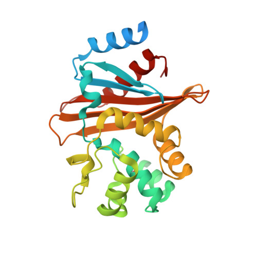 | |
UniProt | |||||
Find proteins for A0A182DW26 (Acinetobacter baumannii (strain ACICU)) Explore A0A182DW26 Go to UniProtKB: A0A182DW26 | |||||
Entity Groups | |||||
| Sequence Clusters | 30% Identity50% Identity70% Identity90% Identity95% Identity100% Identity | ||||
| UniProt Group | A0A182DW26 | ||||
Sequence AnnotationsExpand | |||||
Reference Sequence | |||||
| Modified Residues 1 Unique | |||||
|---|---|---|---|---|---|
| ID | Chains | Type | Formula | 2D Diagram | Parent |
| KCX Query on KCX | A | L-PEPTIDE LINKING | C7 H14 N2 O4 |  | LYS |
| Length ( Å ) | Angle ( ˚ ) |
|---|---|
| a = 37.36 | α = 90 |
| b = 39.79 | β = 91.2 |
| c = 72.09 | γ = 90 |
| Software Name | Purpose |
|---|---|
| REFMAC | refinement |
| SCALA | data reduction |
| HKL-2000 | data scaling |
| MOLREP | phasing |


