4Z9L

THE STRUCTURE OF JNK3 IN COMPLEX WITH AN IMIDAZOLE-PYRIMIDINE INHIBITOR


Experimental Data Snapshot

  • Method: X-RAY DIFFRACTION
  • Resolution: 2.10 Å
  • R-Value Free: 0.226 
  • R-Value Work: 0.200 
  • R-Value Observed: 0.201 

wwPDB Validation   3D Report Full Report


Ligand Structure Quality Assessment 


This is version 1.1 of the entry. See complete history


Literature

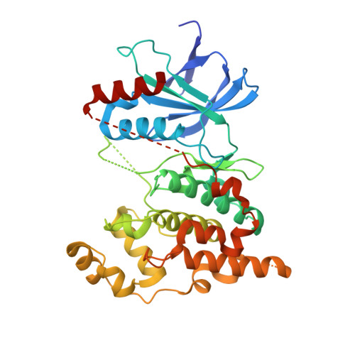
The structure of JNK3 in complex with small molecule inhibitors: structural basis for potency and selectivity.

Scapin, G.Patel, S.B.Lisnock, J.Becker, J.W.LoGrasso, P.V.

(2003) Chem Biol 10: 705-712

  • DOI: https://doi.org/10.1016/s1074-5521(03)00159-5
  • Primary Citation of Related Structures:  
    1PMN, 1PMU, 1PMV, 4Z9L

  • PubMed Abstract: 
  • The c-Jun terminal kinases (JNKs) are members of the mitogen-activated protein (MAP) kinase family and regulate signal transduction in response to environmental stress. Activation of JNK3, a neuronal-specific isoform, has been associated with neurological damage, and as such, JNK3 may represent an attractive target for the treatment of neurological disorders ...

    The c-Jun terminal kinases (JNKs) are members of the mitogen-activated protein (MAP) kinase family and regulate signal transduction in response to environmental stress. Activation of JNK3, a neuronal-specific isoform, has been associated with neurological damage, and as such, JNK3 may represent an attractive target for the treatment of neurological disorders. The MAP kinases share between 50% and 80% sequence identity. In order to obtain efficacious and safe compounds, it is necessary to address the issues of potency and selectivity. We report here four crystal structures of JNK3 in complex with three different classes of inhibitors. These structures provide a clear picture of the interactions that each class of compound made with the kinase. Knowledge of the atomic interactions involved in these diverse binding modes provides a platform for structure-guided modification of these compounds, or the de novo design of novel inhibitors that could satisfy the need for potency and selectivity.


    Related Citations: 
    • Achieving High Quality Ligand Chemistry in Protein-Ligand Crystal Structures for Drug Design
      Smart, O.S., Bricogne, G.
      (2015) Multifaceted Roles Of Crystallography In Modern Drug Discovery --: 165

    Organizational Affiliation

    Department of Medicinal Chemistry, Merck Research Laboratories, P.O. Box 2000, Rahway, NJ 07065, USA. giovanna_scapin@merck.com


Macromolecules
Find similar proteins by:  (by identity cutoff)  |  3D Structure
Entity ID: 1
MoleculeChainsSequence LengthOrganismDetailsImage
Mitogen-activated protein kinase 10364Homo sapiensMutation(s): 0 
Gene Names: MAPK10JNK3JNK3APRKM10SAPK1B
EC: 2.7.11.24
UniProt & NIH Common Fund Data Resources
Find proteins for P53779 (Homo sapiens)
Explore P53779 
Go to UniProtKB:  P53779
PHAROS:  P53779
Entity Groups  
Sequence Clusters30% Identity50% Identity70% Identity90% Identity95% Identity100% Identity
UniProt GroupP53779
Protein Feature View
Expand
  • Reference Sequence
Small Molecules
Ligands 3 Unique
IDChainsName / Formula / InChI Key2D Diagram3D Interactions
880
Query on 880

Download Ideal Coordinates CCD File 
C [auth A]CYCLOHEXYL-{4-[5-(3,4-DICHLOROPHENYL)-2-PIPERIDIN-4-YL-3-PROPYL-3H-IMIDAZOL-4-YL]-PYRIMIDIN-2-YL}AMINE
C27 H34 Cl2 N6
UQFRSHRWRKYNDE-UHFFFAOYSA-N
 Ligand Interaction
ANP
Query on ANP

Download Ideal Coordinates CCD File 
B [auth A]PHOSPHOAMINOPHOSPHONIC ACID-ADENYLATE ESTER
C10 H17 N6 O12 P3
PVKSNHVPLWYQGJ-KQYNXXCUSA-N
 Ligand Interaction
UNX
Query on UNX

Download Ideal Coordinates CCD File 
D [auth A],
E [auth A],
F [auth A]
UNKNOWN ATOM OR ION
X
 Ligand Interaction
Binding Affinity Annotations 
IDSourceBinding Affinity
880 Binding MOAD:  4Z9L IC50: 1.6 (nM) from 1 assay(s)
BindingDB:  4Z9L IC50: min: 1.6, max: 2 (nM) from 3 assay(s)
Experimental Data & Validation

Experimental Data

  • Method: X-RAY DIFFRACTION
  • Resolution: 2.10 Å
  • R-Value Free: 0.226 
  • R-Value Work: 0.200 
  • R-Value Observed: 0.201 
  • Space Group: P 21 21 21
Unit Cell:
Length ( Å )Angle ( ˚ )
a = 50.951α = 90
b = 71.525β = 90
c = 106γ = 90
Software Package:
Software NamePurpose
BUSTERrefinement
HKL-2000data reduction
SCALEPACKdata scaling
Cootmodel building
CNXphasing

Structure Validation

View Full Validation Report


Ligand Structure Quality Assessment 


Entry History 

Deposition Data

Revision History  (Full details and data files)

  • Version 1.0: 2015-05-06
    Type: Initial release
  • Version 1.1: 2023-09-27
    Changes: Advisory, Data collection, Database references, Derived calculations, Refinement description, Source and taxonomy, Structure summary