4Z0L

The murine cyclooxygenase-2 complexed with a nido-dicarbaborate-containing indomethacin derivative


Experimental Data Snapshot

  • Method: X-RAY DIFFRACTION
  • Resolution: 2.29 Å
  • R-Value Free: 0.250 
  • R-Value Work: 0.210 
  • R-Value Observed: 0.212 

wwPDB Validation   3D Report Full Report


Ligand Structure Quality Assessment 


This is version 2.1 of the entry. See complete history


Literature

nido-Dicarbaborate Induces Potent and Selective Inhibition of Cyclooxygenase-2.

Neumann, W.Xu, S.Sarosi, M.B.Scholz, M.S.Crews, B.C.Ghebreselasie, K.Banerjee, S.Marnett, L.J.Hey-Hawkins, E.

(2016) ChemMedChem 11: 175-178

  • DOI: https://doi.org/10.1002/cmdc.201500199
  • Primary Citation of Related Structures:  
    4Z0L

  • PubMed Abstract: 

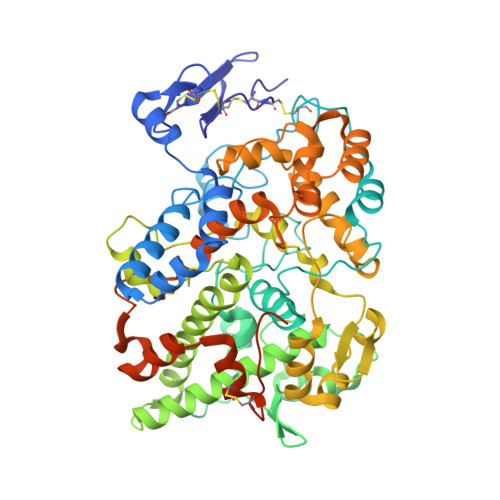
    Carbaboranes are increasingly studied as pharmacophores, particularly as replacements for aromatic systems. However, especially ortho-carbaborane is prone to degradation of the cluster, which hampers biological application. This study demonstrates that deboronation of the cluster may not only lead to a more active analogue, but can also improve the solubility and stability of a carbaborane-containing inhibitor. Notably, introduction of a nido-dicarbaborate cluster into the cyclooxygenase (COX) inhibitor indomethacin results in remarkably increased inhibitory potency and selectivity for COX-2 relative to the respective phenyl analogue. The first crystal structure of a carbaborane-containing inhibitor bound to COX-2 further reveals a novel binding mode for the inhibitor that is strikingly different from that of indomethacin. These results indicate that nido-dicarbaborate is a promising pharmacophore that exhibits properties which are also highly beneficial for its introduction into other inhibitor classes.


  • Organizational Affiliation

    Institut für Anorganische Chemie, Universität Leipzig, Johannisallee 29, 04103, Leipzig, Germany.


Macromolecules
Find similar proteins by:  (by identity cutoff)  |  3D Structure
Entity ID: 1
MoleculeChains Sequence LengthOrganismDetailsImage
Prostaglandin G/H synthase 2
A, B, C, D
587Mus musculusMutation(s): 0 
Gene Names: Ptgs2Cox-2Cox2Pghs-bTis10
EC: 1.14.99.1
UniProt
Find proteins for Q05769 (Mus musculus)
Explore Q05769 
Go to UniProtKB:  Q05769
Entity Groups  
Sequence Clusters30% Identity50% Identity70% Identity90% Identity95% Identity100% Identity
UniProt GroupQ05769
Sequence Annotations
Expand
  • Reference Sequence
Oligosaccharides

Help

Entity ID: 2
MoleculeChains Length2D Diagram Glycosylation3D Interactions
2-acetamido-2-deoxy-beta-D-glucopyranose-(1-4)-2-acetamido-2-deoxy-beta-D-glucopyranose
E, F, G, H
2N-Glycosylation
Glycosylation Resources
GlyTouCan:  G42666HT
GlyCosmos:  G42666HT
GlyGen:  G42666HT
Small Molecules
Ligands 5 Unique
IDChains Name / Formula / InChI Key2D Diagram3D Interactions
HEM
Query on HEM

Download Ideal Coordinates CCD File 
BA [auth D],
I [auth A],
O [auth B],
U [auth C]
PROTOPORPHYRIN IX CONTAINING FE
C34 H32 Fe N4 O4
KABFMIBPWCXCRK-RGGAHWMASA-L
N1B
Query on N1B

Download Ideal Coordinates CCD File 
FA [auth D],
M [auth A],
S [auth B],
Y [auth C]
(S)-7-{[5-methoxy-2-methyl-3-(methoxycarbonylmethyl)-1H-indolyl]carbonyl}-7,8-dicarba-nido-dodeca-hydroundecaborate
C16 H14 B9 N O4
FOWIJUKMQDHTEE-UHFFFAOYSA-N
4LA
Query on 4LA

Download Ideal Coordinates CCD File 
EA [auth D],
L [auth A],
R [auth B],
X [auth C]
(R)-7-{[5-methoxy-2-methyl-3-(methoxycarbonylmethyl)-1H-indolyl]carbonyl}-7,8-dicarba-nido-dodeca-hydroundecaborate
C16 H14 B9 N O4
FOWIJUKMQDHTEE-UHFFFAOYSA-N
BOG
Query on BOG

Download Ideal Coordinates CCD File 
AA [auth C],
GA [auth D],
N [auth A],
T [auth B],
Z [auth C]
octyl beta-D-glucopyranoside
C14 H28 O6
HEGSGKPQLMEBJL-RKQHYHRCSA-N
NAG
Query on NAG

Download Ideal Coordinates CCD File 
CA [auth D]
DA [auth D]
J [auth A]
K [auth A]
P [auth B]
CA [auth D],
DA [auth D],
J [auth A],
K [auth A],
P [auth B],
Q [auth B],
V [auth C],
W [auth C]
2-acetamido-2-deoxy-beta-D-glucopyranose
C8 H15 N O6
OVRNDRQMDRJTHS-FMDGEEDCSA-N
Binding Affinity Annotations 
IDSourceBinding Affinity
N1B Binding MOAD:  4Z0L IC50: 51 (nM) from 1 assay(s)
4LA Binding MOAD:  4Z0L IC50: 51 (nM) from 1 assay(s)
Experimental Data & Validation

Experimental Data

  • Method: X-RAY DIFFRACTION
  • Resolution: 2.29 Å
  • R-Value Free: 0.250 
  • R-Value Work: 0.210 
  • R-Value Observed: 0.212 
  • Space Group: P 21 21 2
Unit Cell:
Length ( Å )Angle ( ˚ )
a = 180.944α = 90
b = 135.011β = 90
c = 124.195γ = 90
Software Package:
Software NamePurpose
PHENIXrefinement
XDSdata reduction
XDSdata scaling
PHASERphasing

Structure Validation

View Full Validation Report


Ligand Structure Quality Assessment 


Entry History & Funding Information

Deposition Data


Funding OrganizationLocationGrant Number
National Institutes of Health/National Cancer Institute (NIH/NCI)United StatesCA89450
German Research Foundation (DFG)Germany--
the Fonds der Chemischen IndustrieGermany--
the Free State of SaxonyGermanyESF-NFG 100148835
National Institutes of Health/National Institute of General Medical Sciences (NIH/NIGMS)United StatesP41 GM103403

Revision History  (Full details and data files)

  • Version 1.0: 2015-06-10
    Type: Initial release
  • Version 1.1: 2015-07-01
    Changes: Database references
  • Version 1.2: 2016-02-03
    Changes: Database references
  • Version 1.3: 2017-08-23
    Changes: Data collection, Derived calculations, Refinement description
  • Version 1.4: 2017-09-06
    Changes: Author supporting evidence
  • Version 1.5: 2017-09-27
    Changes: Author supporting evidence
  • Version 1.6: 2019-12-04
    Changes: Author supporting evidence
  • Version 2.0: 2020-07-29
    Type: Remediation
    Reason: Carbohydrate remediation
    Changes: Atomic model, Data collection, Derived calculations, Structure summary
  • Version 2.1: 2023-09-27
    Changes: Data collection, Database references, Refinement description, Structure summary