4YO3

Enteroaggregative Escherichia Coli TssA N-terminal fragment


Experimental Data Snapshot

  • Method: X-RAY DIFFRACTION
  • Resolution: 3.37 Å
  • R-Value Free: 0.261 
  • R-Value Work: 0.250 
  • R-Value Observed: 0.251 

wwPDB Validation   3D Report Full Report


This is version 1.2 of the entry. See complete history


Literature

Priming and polymerization of a bacterial contractile tail structure.

Zoued, A.Durand, E.Brunet, Y.R.Spinelli, S.Douzi, B.Guzzo, M.Flaugnatti, N.Legrand, P.Journet, L.Fronzes, R.Mignot, T.Cambillau, C.Cascales, E.

(2016) Nature 531: 47-52

  • DOI: https://doi.org/10.1038/nature17182
  • Primary Citation of Related Structures:  
    4YO3, 4YO5

  • PubMed Abstract: 

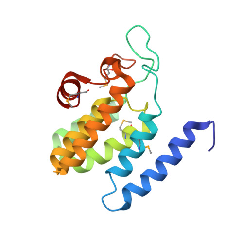
    Contractile tails are composed of an inner tube wrapped by an outer sheath assembled in an extended, metastable conformation that stores mechanical energy necessary for its contraction. Contraction is used to propel the rigid inner tube towards target cells for DNA or toxin delivery. Although recent studies have revealed the structure of the contractile sheath of the type VI secretion system, the mechanisms by which its polymerization is controlled and coordinated with the assembly of the inner tube remain unknown. Here we show that the starfish-like TssA dodecameric complex interacts with tube and sheath components. Fluorescence microscopy experiments in enteroaggregative Escherichia coli reveal that TssA binds first to the type VI secretion system membrane core complex and then initiates tail polymerization. TssA remains at the tip of the growing structure and incorporates new tube and sheath blocks. On the basis of these results, we propose that TssA primes and coordinates tail tube and sheath biogenesis.


  • Organizational Affiliation

    Laboratoire d'Ingénierie des Systèmes Macromoléculaires, Institut de Microbiologie de la Méditerranée, CNRS UMR7255, Aix-Marseille Université, 31 Chemin Joseph Aiguier, 13402 Marseille Cedex 20, France.


Macromolecules
Find similar proteins by:  (by identity cutoff)  |  3D Structure
Entity ID: 1
MoleculeChains Sequence LengthOrganismDetailsImage
TssA
A, B, C, D, E
A, B, C, D, E, F, G, H, I, J, K, L
157Escherichia coli 042Mutation(s): 0 
Gene Names: EC55989_3324
UniProt
Find proteins for D3GUX1 (Escherichia coli O44:H18 (strain 042 / EAEC))
Explore D3GUX1 
Go to UniProtKB:  D3GUX1
Entity Groups  
Sequence Clusters30% Identity50% Identity70% Identity90% Identity95% Identity100% Identity
UniProt GroupD3GUX1
Sequence Annotations
Expand
  • Reference Sequence
Small Molecules
Modified Residues  1 Unique
IDChains TypeFormula2D DiagramParent
MSE
Query on MSE
A, B, C, D, E
A, B, C, D, E, F, G, H, I, J, K, L
L-PEPTIDE LINKINGC5 H11 N O2 SeMET
Experimental Data & Validation

Experimental Data

  • Method: X-RAY DIFFRACTION
  • Resolution: 3.37 Å
  • R-Value Free: 0.261 
  • R-Value Work: 0.250 
  • R-Value Observed: 0.251 
  • Space Group: P 21 21 2
Unit Cell:
Length ( Å )Angle ( ˚ )
a = 134.13α = 90
b = 178.05β = 90
c = 106.76γ = 90
Software Package:
Software NamePurpose
BUSTERrefinement
XDSdata reduction
SCALAdata scaling
SHELXDEphasing

Structure Validation

View Full Validation Report


Entry History & Funding Information

Deposition Data


Funding OrganizationLocationGrant Number
French National Research AgencyFranceANR-10-JCJC-1303-03
Fondation pour la Recherche MedicaleFranceFRM DEQ2011-0421282
Fondation pour la Recherche MedicaleFranceSPF20101221116

Revision History  (Full details and data files)

  • Version 1.0: 2016-02-17
    Type: Initial release
  • Version 1.1: 2016-03-09
    Changes: Database references
  • Version 1.2: 2017-08-30
    Changes: Author supporting evidence