Structural basis for 2'-5'-oligoadenylate binding and enzyme activity of a viral RNase L antagonist.
Ogden, K.M., Hu, L., Jha, B.K., Sankaran, B., Weiss, S.R., Silverman, R.H., Patton, J.T., Prasad, B.V.(2015) J Virol 89: 6633-6645
- PubMed: 25878106 Search on PubMedSearch on PubMed Central
- DOI: https://doi.org/10.1128/JVI.00701-15
- Primary Citation Related Structures:
4RPT, 4YE2 - PubMed Abstract:
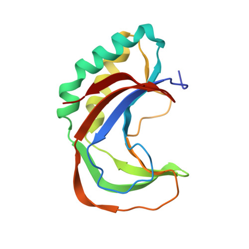
Synthesis of 2'-5'-oligoadenylates (2-5A) by oligoadenylate synthetase (OAS) is an important innate cellular response that limits viral replication by activating the latent cellular RNase, RNase L, to degrade single-stranded RNA. Some rotaviruses and coronaviruses antagonize the OAS/RNase L pathway through the activity of an encoded 2H phosphoesterase domain that cleaves 2-5A. These viral 2H phosphoesterases are phylogenetically related to the cellular A kinase anchoring protein 7 (AKAP7) and share a core structure and an active site that contains two well-defined HΦ(S/T)Φ (where Φ is a hydrophobic residue) motifs, but their mechanism of substrate binding is unknown. Here, we report the structures of a viral 2H phosphoesterase, the C-terminal domain (CTD) of the group A rotavirus (RVA) VP3 protein, both alone and in complex with 2-5A. The domain forms a compact fold, with a concave β-sheet that contains the catalytic cleft, but it lacks two α-helical regions and two β-strands observed in AKAP7 and other 2H phosphoesterases. The cocrystal structure shows significant conformational changes in the R loop upon ligand binding. Bioinformatics and biochemical analyses reveal that conserved residues and residues required for catalytic activity and substrate binding comprise the catalytic motifs and a region on one side of the binding cleft. We demonstrate that the VP3 CTD of group B rotavirus, but not that of group G, cleaves 2-5A. These findings suggest that the VP3 CTD is a streamlined version of a 2H phosphoesterase with a ligand-binding mechanism that is shared among 2H phosphodiesterases that cleave 2-5A. The C-terminal domain (CTD) of rotavirus VP3 is a 2H phosphoesterase that cleaves 2'-5'-oligoadenylates (2-5A), potent activators of an important innate cellular antiviral pathway. 2H phosphoesterase superfamily proteins contain two conserved catalytic motifs and a proposed core structure. Here, we present structures of a viral 2H phosphoesterase, the rotavirus VP3 CTD, alone and in complex with its substrate, 2-5A. The domain lacks two α-helical regions and β-strands present in other 2H phosphoesterases. A loop of the protein undergoes significant structural changes upon substrate binding. Together with our bioinformatics and biochemical findings, the crystal structures suggest that the RVA VP3 CTD domain is a streamlined version of a cellular enzyme that shares a ligand-binding mechanism with other 2H phosphodiesterases that cleave 2-5A but differs from those of 2H phosphodiesterases that cleave other substrates. These findings may aid in the future design of antivirals targeting viral phosphodiesterases with cleavage specificity for 2-5A.