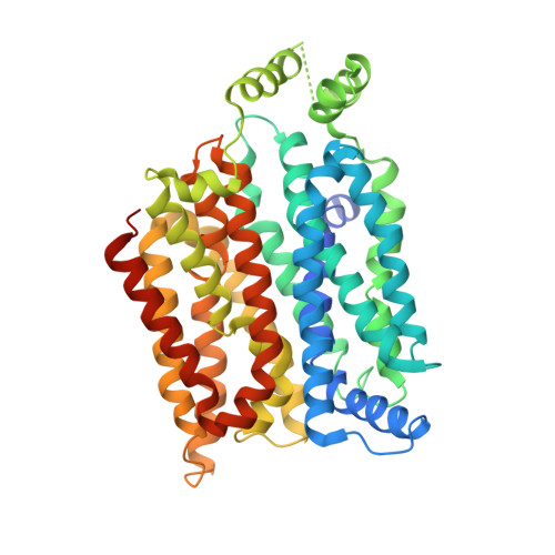
4YB9

Crystal structure of the Bovine Fructose transporter GLUT5 in an open inward-facing conformation


Experimental Data Snapshot

  • Method: X-RAY DIFFRACTION
  • Resolution: 3.20 Å
  • R-Value Free: 0.258 
  • R-Value Work: 0.237 
  • R-Value Observed: 0.239 

Starting Model: experimental
View more details

wwPDB Validation   3D Report Full Report


This is version 1.3 of the entry. See complete history


Literature

Structure and mechanism of the mammalian fructose transporter GLUT5.

Nomura, N.Verdon, G.Kang, H.J.Shimamura, T.Nomura, Y.Sonoda, Y.Hussien, S.A.Qureshi, A.A.Coincon, M.Sato, Y.Abe, H.Nakada-Nakura, Y.Hino, T.Arakawa, T.Kusano-Arai, O.Iwanari, H.Murata, T.Kobayashi, T.Hamakubo, T.Kasahara, M.Iwata, S.Drew, D.

(2015) Nature 526: 397-401

  • DOI: https://doi.org/10.1038/nature14909
  • Primary Citation of Related Structures:  
    4YB9, 4YBQ

  • PubMed Abstract: 

    The altered activity of the fructose transporter GLUT5, an isoform of the facilitated-diffusion glucose transporter family, has been linked to disorders such as type 2 diabetes and obesity. GLUT5 is also overexpressed in certain tumour cells, and inhibitors are potential drugs for these conditions. Here we describe the crystal structures of GLUT5 from Rattus norvegicus and Bos taurus in open outward- and open inward-facing conformations, respectively. GLUT5 has a major facilitator superfamily fold like other homologous monosaccharide transporters. On the basis of a comparison of the inward-facing structures of GLUT5 and human GLUT1, a ubiquitous glucose transporter, we show that a single point mutation is enough to switch the substrate-binding preference of GLUT5 from fructose to glucose. A comparison of the substrate-free structures of GLUT5 with occluded substrate-bound structures of Escherichia coli XylE suggests that, in addition to global rocker-switch-like re-orientation of the bundles, local asymmetric rearrangements of carboxy-terminal transmembrane bundle helices TM7 and TM10 underlie a 'gated-pore' transport mechanism in such monosaccharide transporters.


  • Organizational Affiliation

    Department of Cell Biology, Graduate School of Medicine, Kyoto University, Konoe-cho, Yoshida, Sakyo-ku, Kyoto 606-8501, Japan.


Macromolecules
Find similar proteins by:  (by identity cutoff)  |  3D Structure
Entity ID: 1
MoleculeChains Sequence LengthOrganismDetailsImage
Solute carrier family 2, facilitated glucose transporter member 5A [auth D]479Bos taurusMutation(s): 1 
Gene Names: SLC2A5GLUT5
Membrane Entity: Yes 
UniProt
Find proteins for P58353 (Bos taurus)
Explore P58353 
Go to UniProtKB:  P58353
Entity Groups  
Sequence Clusters30% Identity50% Identity70% Identity90% Identity95% Identity100% Identity
UniProt GroupP58353
Sequence Annotations
Expand
  • Reference Sequence
Experimental Data & Validation

Experimental Data

  • Method: X-RAY DIFFRACTION
  • Resolution: 3.20 Å
  • R-Value Free: 0.258 
  • R-Value Work: 0.237 
  • R-Value Observed: 0.239 
  • Space Group: P 21 21 21
Unit Cell:
Length ( Å )Angle ( ˚ )
a = 74.61α = 90
b = 112.15β = 90
c = 139.57γ = 90
Software Package:
Software NamePurpose
PHENIXrefinement
HKL-2000data scaling
HKL-2000data reduction
PHASERphasing

Structure Validation

View Full Validation Report


Entry History & Funding Information

Deposition Data


Funding OrganizationLocationGrant Number
Biotechnology and Biological Sciences Research CouncilUnited KingdomBB/G02325/1
Wellcome TrustUnited Kingdom062164/Z/00/Z

Revision History  (Full details and data files)

  • Version 1.0: 2015-10-14
    Type: Initial release
  • Version 1.1: 2015-10-21
    Changes: Database references
  • Version 1.2: 2017-08-30
    Changes: Author supporting evidence
  • Version 1.3: 2024-05-01
    Changes: Data collection, Database references, Refinement description