4XW7

Crystal structure of the ZMP riboswitch at 2.50 angstrom


Experimental Data Snapshot

  • Method: X-RAY DIFFRACTION
  • Resolution: 2.50 Å
  • R-Value Free: 0.274 
  • R-Value Work: 0.249 
  • R-Value Observed: 0.252 

wwPDB Validation   3D Report Full Report


Ligand Structure Quality Assessment 


This is version 1.3 of the entry. See complete history


Literature

Metal Ion-Mediated Nucleobase Recognition by the ZTP Riboswitch.

Trausch, J.J.Marcano-Velazquez, J.G.Matyjasik, M.M.Batey, R.T.

(2015) Chem Biol 22: 829-837

  • DOI: https://doi.org/10.1016/j.chembiol.2015.06.007
  • Primary Citation of Related Structures:  
    4XW7, 4XWF

  • PubMed Abstract: 

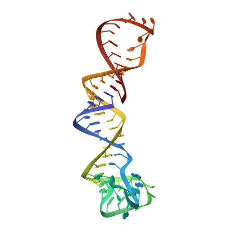
    The ZTP riboswitch is a widespread family of regulatory RNAs that upregulate de novo purine synthesis in response to increased intracellular levels of ZTP or ZMP. As an important intermediate in purine biosynthesis, ZMP also serves as a proxy for the concentration of N10-formyl-tetrahydrofolate, a key component of one-carbon metabolism. Here, we report the structure of the ZTP riboswitch bound to ZMP at a resolution of 1.80 Å. The RNA contains two subdomains brought together through a long-range pseudoknot further stabilized through helix-helix packing. ZMP is bound at the subdomain interface of the RNA through a set of interactions with the base, ribose sugar, and phosphate moieties of the ligand. Unique to nucleobase recognition by RNAs, the Z base is inner-sphere coordinated to a magnesium cation bound by two backbone phosphates. This interaction, along with steric hindrance by the backbone, imparts specificity over chemically similar compounds such as ATP/AMP.


  • Organizational Affiliation

    Department of Chemistry and Biochemistry, University of Colorado at Boulder, Campus Box 596, Boulder, CO 80309-0596, USA.


Macromolecules
Find similar nucleic acids by:  (by identity cutoff)  |  3D Structure
Entity ID: 1
MoleculeChains LengthOrganismImage
pfl RNA64Schaalia odontolytica
Sequence Annotations
Expand
  • Reference Sequence
Small Molecules
Ligands 3 Unique
IDChains Name / Formula / InChI Key2D Diagram3D Interactions
AMZ
Query on AMZ

Download Ideal Coordinates CCD File 
B [auth A]AMINOIMIDAZOLE 4-CARBOXAMIDE RIBONUCLEOTIDE
C9 H15 N4 O8 P
NOTGFIUVDGNKRI-UUOKFMHZSA-N
IRI
Query on IRI

Download Ideal Coordinates CCD File 
P [auth A],
Q [auth A],
R [auth A],
S [auth A]
IRIDIUM HEXAMMINE ION
H18 Ir N6
CGMAOQLDNKCXGK-UHFFFAOYSA-N
MG
Query on MG

Download Ideal Coordinates CCD File 
C [auth A]
D [auth A]
E [auth A]
F [auth A]
G [auth A]
C [auth A],
D [auth A],
E [auth A],
F [auth A],
G [auth A],
H [auth A],
I [auth A],
J [auth A],
K [auth A],
L [auth A],
M [auth A],
N [auth A],
O [auth A]
MAGNESIUM ION
Mg
JLVVSXFLKOJNIY-UHFFFAOYSA-N
Experimental Data & Validation

Experimental Data

  • Method: X-RAY DIFFRACTION
  • Resolution: 2.50 Å
  • R-Value Free: 0.274 
  • R-Value Work: 0.249 
  • R-Value Observed: 0.252 
  • Space Group: P 43 21 2
Unit Cell:
Length ( Å )Angle ( ˚ )
a = 40.16α = 90
b = 40.16β = 90
c = 234.91γ = 90
Software Package:
Software NamePurpose
PHENIXrefinement
CrystalCleardata collection
MOSFLMdata reduction
PHASERphasing

Structure Validation

View Full Validation Report


Ligand Structure Quality Assessment 


Entry History & Funding Information

Deposition Data


Funding OrganizationLocationGrant Number
National Institutes of Health/National Institute of General Medical Sciences (NIH/NIGMS)United StatesGM073850

Revision History  (Full details and data files)

  • Version 1.0: 2015-09-09
    Type: Initial release
  • Version 1.1: 2017-09-27
    Changes: Author supporting evidence, Database references, Derived calculations, Refinement description
  • Version 1.2: 2019-12-25
    Changes: Author supporting evidence
  • Version 1.3: 2024-02-28
    Changes: Data collection, Database references, Derived calculations