Elongation of the Poly-gamma-glutamate Tail of F420 Requires Both Domains of the F420: gamma-Glutamyl Ligase (FbiB) of Mycobacterium tuberculosis.
Bashiri, G., Rehan, A.M., Sreebhavan, S., Baker, H.M., Baker, E.N., Squire, C.J.(2016) J Biol Chem 291: 6882-6894
- PubMed: 26861878 Search on PubMedSearch on PubMed Central
- DOI: https://doi.org/10.1074/jbc.M115.689026
- Primary Citation Related Structures:
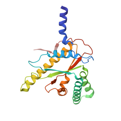
4XOM, 4XOO, 4XOQ - PubMed Abstract:
Cofactor F420is an electron carrier with a major role in the oxidoreductive reactions ofMycobacterium tuberculosis, the causative agent of tuberculosis. A γ-glutamyl ligase catalyzes the final steps of the F420biosynthesis pathway by successive additions ofl-glutamate residues to F420-0, producing a poly-γ-glutamate tail. The enzyme responsible for this reaction in archaea (CofE) comprises a single domain and produces F420-2 as the major species. The homologousM. tuberculosisenzyme, FbiB, is a two-domain protein and produces F420with predominantly 5-7l-glutamate residues in the poly-γ-glutamate tail. The N-terminal domain of FbiB is homologous to CofE with an annotated γ-glutamyl ligase activity, whereas the C-terminal domain has sequence similarity to an FMN-dependent family of nitroreductase enzymes. Here we demonstrate that full-length FbiB adds multiplel-glutamate residues to F420-0in vitroto produce F420-5 after 24 h; communication between the two domains is critical for full γ-glutamyl ligase activity. We also present crystal structures of the C-terminal domain of FbiB in apo-, F420-0-, and FMN-bound states, displaying distinct sites for F420-0 and FMN ligands that partially overlap. Finally, we discuss the features of a full-length structural model produced by small angle x-ray scattering and its implications for the role of N- and C-terminal domains in catalysis.
- From the Structural Biology Laboratory, School of Biological Sciences and Maurice Wilkins Centre for Molecular Biodiscovery, and.
Organizational Affiliation: