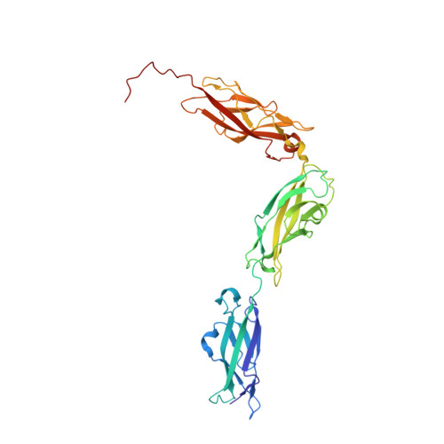
4XHZ

Crystal Structure of Human Protocadherin-15 EC8-10


Experimental Data Snapshot

  • Method: X-RAY DIFFRACTION
  • Resolution: 2.80 Å
  • R-Value Free: 0.200 
  • R-Value Work: 0.170 
  • R-Value Observed: 0.171 

wwPDB Validation   3D Report Full Report


This is version 1.5 of the entry. See complete history


Literature

An elastic element in the protocadherin-15 tip link of the inner ear.

Araya-Secchi, R.Neel, B.L.Sotomayor, M.

(2016) Nat Commun 7: 13458-13458

  • DOI: https://doi.org/10.1038/ncomms13458
  • Primary Citation of Related Structures:  
    4XHZ, 5KJ4

  • PubMed Abstract: 

    Tip link filaments convey force and gate inner-ear hair-cell transduction channels to mediate perception of sound and head movements. Cadherin-23 and protocadherin-15 form tip links through a calcium-dependent interaction of their extracellular domains made of multiple extracellular cadherin (EC) repeats. These repeats are structurally similar, but not identical in sequence, often featuring linkers with conserved calcium-binding sites that confer mechanical strength to them. Here we present the X-ray crystal structures of human protocadherin-15 EC8-EC10 and mouse EC9-EC10, which show an EC8-9 canonical-like calcium-binding linker, and an EC9-10 calcium-free linker that alters the linear arrangement of EC repeats. Molecular dynamics simulations and small-angle X-ray scattering experiments support this non-linear conformation. Simulations also suggest that unbending of EC9-10 confers some elasticity to otherwise rigid tip links. The new structure provides a first view of protocadherin-15's non-canonical EC linkers and suggests how they may function in inner-ear mechanotransduction, with implications for other cadherins.


  • Organizational Affiliation

    Department of Chemistry and Biochemistry, The Ohio State University, 484 W 12th Avenue, Columbus, Ohio 43210, USA.


Macromolecules
Find similar proteins by:  (by identity cutoff)  |  3D Structure
Entity ID: 1
MoleculeChains Sequence LengthOrganismDetailsImage
Protocadherin-15338Homo sapiensMutation(s): 0 
Gene Names: PCDH15USH1F
UniProt & NIH Common Fund Data Resources
Find proteins for Q96QU1 (Homo sapiens)
Explore Q96QU1 
Go to UniProtKB:  Q96QU1
PHAROS:  Q96QU1
GTEx:  ENSG00000150275 
Entity Groups  
Sequence Clusters30% Identity50% Identity70% Identity90% Identity95% Identity100% Identity
UniProt GroupQ96QU1
Sequence Annotations
Expand
  • Reference Sequence
Experimental Data & Validation

Experimental Data

  • Method: X-RAY DIFFRACTION
  • Resolution: 2.80 Å
  • R-Value Free: 0.200 
  • R-Value Work: 0.170 
  • R-Value Observed: 0.171 
  • Space Group: P 63 2 2
Unit Cell:
Length ( Å )Angle ( ˚ )
a = 157.929α = 90
b = 157.929β = 90
c = 142.898γ = 120
Software Package:
Software NamePurpose
REFMACrefinement
HKL-2000data reduction
HKL-2000data scaling
PHASERphasing

Structure Validation

View Full Validation Report


Entry History & Funding Information

Deposition Data


Funding OrganizationLocationGrant Number
National Institutes of Health/National Institute on Deafness and Other Communication Disorders (NIH/NIDCD)United StatesK99/R00 DC012534

Revision History  (Full details and data files)

  • Version 1.0: 2016-09-21
    Type: Initial release
  • Version 1.1: 2016-11-23
    Changes: Database references
  • Version 1.2: 2016-12-21
    Changes: Database references
  • Version 1.3: 2017-09-06
    Changes: Author supporting evidence
  • Version 1.4: 2019-12-18
    Changes: Author supporting evidence
  • Version 1.5: 2023-09-27
    Changes: Data collection, Database references, Derived calculations, Refinement description