


Crystal Structure of Dimeric Human Peroxiredoxin-1 C83S Mutant
Cho, K.J., Park, Y., Khan, T.G., Lee, J.-H., Kim, S., Seok, J.H., Chung, Y.B., Cho, A.E., Choi, Y., Chang, T.-S., Kim, K.H.(2015) Bull Korean Chem Soc 36: 1543-1545
Experimental Data Snapshot
Starting Model: experimental
View more details
(2015) Bull Korean Chem Soc 36: 1543-1545
Entity ID: 1 | |||||
|---|---|---|---|---|---|
| Molecule | Chains | Sequence Length | Organism | Details | Image |
| Peroxiredoxin-1 | 219 | Homo sapiens | Mutation(s): 1 Gene Names: PRDX1, PAGA, PAGB, TDPX2 EC: 1.11.1.15 (PDB Primary Data), 1.11.1.24 (UniProt) | 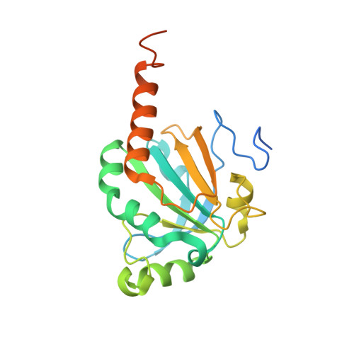 | |
UniProt & NIH Common Fund Data Resources | |||||
PHAROS: Q06830 GTEx: ENSG00000117450 | |||||
Entity Groups | |||||
| Sequence Clusters | 30% Identity50% Identity70% Identity90% Identity95% Identity100% Identity | ||||
| UniProt Group | Q06830 | ||||
Sequence AnnotationsExpand | |||||
Reference Sequence | |||||
| Ligands 2 Unique | |||||
|---|---|---|---|---|---|
| ID | Chains | Name / Formula / InChI Key | 2D Diagram | 3D Interactions | |
| CPS Download:Ideal Coordinates CCD File | H [auth A], J [auth D], K [auth E] | 3-[(3-CHOLAMIDOPROPYL)DIMETHYLAMMONIO]-1-PROPANESULFONATE C32 H58 N2 O7 S UMCMPZBLKLEWAF-BCTGSCMUSA-N |  | ||
| GOL Download:Ideal Coordinates CCD File | G [auth A], I [auth C], L [auth F] | GLYCEROL C3 H8 O3 PEDCQBHIVMGVHV-UHFFFAOYSA-N |  | ||
| Length ( Å ) | Angle ( ˚ ) |
|---|---|
| a = 138.788 | α = 90 |
| b = 80.128 | β = 89.99 |
| c = 125.618 | γ = 90 |
| Software Name | Purpose |
|---|---|
| REFMAC | refinement |
| HKL-2000 | data reduction |
| PDB_EXTRACT | data extraction |
| HKL-2000 | data scaling |
| MOLREP | phasing |


