4WVI

Crystal structure of the Type-I signal peptidase from Staphylococcus aureus (SpsB) in complex with a substrate peptide (pep2).


Experimental Data Snapshot

  • Method: X-RAY DIFFRACTION
  • Resolution: 1.90 Å
  • R-Value Free: 0.219 
  • R-Value Work: 0.188 
  • R-Value Observed: 0.190 

wwPDB Validation   3D Report Full Report


This is version 2.0 of the entry. See complete history


Literature

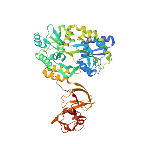
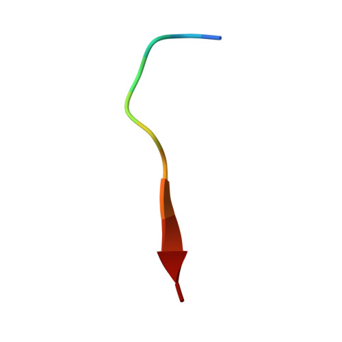
Peptide binding to a bacterial signal peptidase visualized by peptide tethering and carrier-driven crystallization.

Ting, Y.T.Harris, P.W.Batot, G.Brimble, M.A.Baker, E.N.Young, P.G.

(2016) IUCrJ 3: 10-19

  • DOI: 10.1107/S2052252515019971
  • Primary Citation of Related Structures:  
    4WVG, 4WVH, 4WVI, 4WVJ

  • PubMed Abstract: 
  • Bacterial type I signal peptidases (SPases) are membrane-anchored serine proteases that process the signal peptides of proteins exported via the Sec and Tat secretion systems. Despite their crucial importance for bacterial virulence and their attractiveness as drug targets, only one such enzyme, LepB from Escherichia coli, has been structurally characterized, and the transient nature of peptide binding has stymied attempts to directly visualize SPase-substrate complexes ...

    Bacterial type I signal peptidases (SPases) are membrane-anchored serine proteases that process the signal peptides of proteins exported via the Sec and Tat secretion systems. Despite their crucial importance for bacterial virulence and their attractiveness as drug targets, only one such enzyme, LepB from Escherichia coli, has been structurally characterized, and the transient nature of peptide binding has stymied attempts to directly visualize SPase-substrate complexes. Here, the crystal structure of SpsB, the type I signal peptidase from the Gram-positive pathogen Staphylococcus aureus, is reported, and a peptide-tethering strategy that exploits the use of carrier-driven crystallization is described. This enabled the determination of the crystal structures of three SpsB-peptide complexes, both with cleavable substrates and with an inhibitory peptide. SpsB-peptide interactions in these complexes are almost exclusively limited to the canonical signal-peptide motif Ala-X-Ala, for which clear specificity pockets are found. Minimal contacts are made outside this core, with the variable side chains of the peptides accommodated in shallow grooves or exposed faces. These results illustrate how high fidelity is retained despite broad sequence diversity, in a process that is vital for cell survival.


    Organizational Affiliation

    School of Biological Sciences, The University of Auckland, Auckland 1142, New Zealand; Maurice Wilkins Centre for Molecular Biodiscovery, The University of Auckland, Auckland 1142, New Zealand.


Macromolecules
Find similar proteins by:  (by identity cutoff)  |  3D Structure
Entity ID: 1
MoleculeChainsSequence LengthOrganismDetailsImage
Maltose-binding periplasmic protein,Signal peptidase IBA533Escherichia coli K-12Staphylococcus aureus subsp. aureus str. Newman
This entity is chimeric
Mutation(s): 2 
Gene Names: malEZ5632ECs5017spsBSACOL0969
EC: 3.4.21.89
Membrane Entity: Yes 
UniProt
Find proteins for P0AEY0 (Escherichia coli O157:H7)
Explore P0AEY0 
Go to UniProtKB:  P0AEY0
Find proteins for Q5HHB9 (Staphylococcus aureus (strain COL))
Explore Q5HHB9 
Go to UniProtKB:  Q5HHB9
Entity Groups  
Sequence Clusters30% Identity50% Identity70% Identity90% Identity95% Identity100% Identity
UniProt GroupsQ5HHB9P0AEY0
Protein Feature View
Expand
  • Reference Sequence

Find similar proteins by:  Sequence   |   3D Structure  

Entity ID: 2
MoleculeChainsSequence LengthOrganismDetailsImage
substrate peptide (pep2)B [auth D]11Staphylococcus aureusMutation(s): 0 
Entity Groups  
Sequence Clusters30% Identity50% Identity70% Identity90% Identity95% Identity100% Identity
Protein Feature View
Expand
  • Reference Sequence
Oligosaccharides

Help

Entity ID: 3
MoleculeChainsChain Length2D DiagramGlycosylation3D Interactions
alpha-D-glucopyranose-(1-4)-alpha-D-glucopyranoseC [auth B]2N/A Oligosaccharides Interaction
Glycosylation Resources
GlyTouCan:  G07411ON
GlyCosmos:  G07411ON
Biologically Interesting Molecules (External Reference) 1 Unique
Entity ID: 3
IDChainsNameType/Class2D Diagram3D Interactions
PRD_900001
Query on PRD_900001
C [auth B]alpha-maltoseOligosaccharide / Nutrient Ligand Interaction
Experimental Data & Validation

Experimental Data

  • Method: X-RAY DIFFRACTION
  • Resolution: 1.90 Å
  • R-Value Free: 0.219 
  • R-Value Work: 0.188 
  • R-Value Observed: 0.190 
  • Space Group: P 21 21 21
Unit Cell:
Length ( Å )Angle ( ˚ )
a = 63.989α = 90
b = 80.232β = 90
c = 119.879γ = 90
Software Package:
Software NamePurpose
XDSdata scaling
Aimlessdata scaling
PHASERphasing
REFMACrefinement
PDB_EXTRACTdata extraction

Structure Validation

View Full Validation Report


Entry History & Funding Information

Deposition Data


Funding OrganizationLocationGrant Number
Health Research Council (HRC)New Zealand--

Revision History  (Full details and data files)

  • Version 1.0: 2015-09-23
    Type: Initial release
  • Version 1.1: 2016-02-17
    Changes: Database references
  • Version 1.2: 2017-09-20
    Changes: Author supporting evidence, Data collection, Derived calculations, Refinement description
  • Version 1.3: 2018-04-25
    Changes: Data collection, Database references
  • Version 1.4: 2018-06-13
    Changes: Data collection
  • Version 1.5: 2019-01-23
    Changes: Data collection, Source and taxonomy
  • Version 1.6: 2020-01-01
    Changes: Author supporting evidence
  • Version 2.0: 2020-07-29
    Type: Remediation
    Reason: Carbohydrate remediation
    Changes: Atomic model, Data collection, Derived calculations, Non-polymer description, Structure summary