4WUZ | pdb_00004wuz

Crystal structure of lambda exonuclease in complex with DNA and Ca2+


Experimental Data Snapshot

  • Method: X-RAY DIFFRACTION
  • Resolution: 2.38 Å
  • R-Value Free: 
    0.319 (Depositor), 0.314 (DCC) 
  • R-Value Work: 
    0.235 (Depositor), 0.230 (DCC) 
  • R-Value Observed: 
    0.239 (Depositor) 

wwPDB Validation 3D Report Full Report

Validation slider image for 4WUZ

This is version 1.6 of the entry. See complete history

Literature

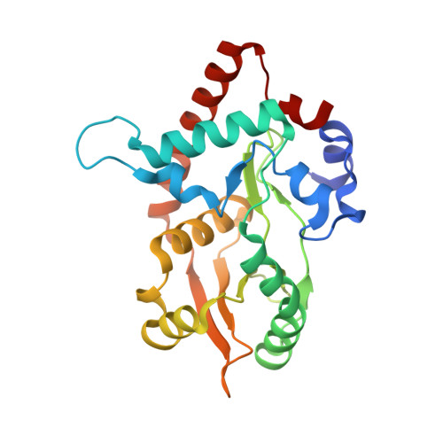
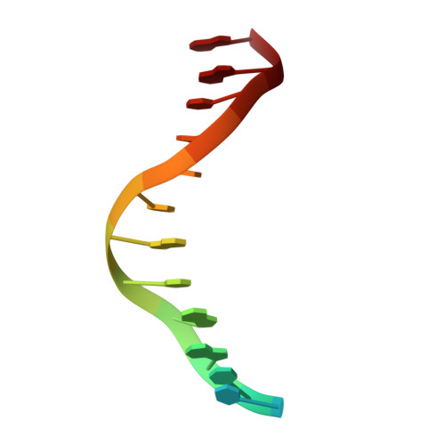
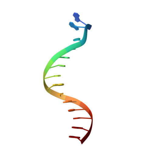
Crystal Structure of lambda Exonuclease in Complex with DNA and Ca(2+).

Zhang, J.Pan, X.Bell, C.E.

(2014) Biochemistry 53: 7415-7425

  • DOI: https://doi.org/10.1021/bi501155q
  • Primary Citation Related Structures: 
    4WUZ

  • PubMed Abstract: 

    Bacteriophage λ exonuclease (λexo) is a ring-shaped homotrimer that resects double-stranded DNA ends in the 5'-3' direction to generate a long 3'-overhang that is a substrate for recombination. λexo is a member of the type II restriction endonuclease-like superfamily of proteins that use a Mg(2+)-dependent mechanism for nucleotide cleavage. A previous structure of λexo in complex with DNA and Mg(2+) was determined using a nuclease defective K131A variant to trap a stable complex. This structure revealed the detailed coordination of the two active site Mg(2+) ions but did not show the interactions involving the side chain of the conserved active site Lys-131 residue. Here, we have determined the crystal structure of wild-type (WT) λexo in complex with the same DNA substrate, but in the presence of Ca(2+) instead of Mg(2+). Surprisingly, there is only one Ca(2+) bound in the active site, near the position of Mg(A) in the structure with Mg(2+). The scissile phosphate is displaced by 2.2 Å relative to its position in the structure with Mg(2+), and the network of interactions involving the attacking water molecule is broken. Thus, the structure does not represent a catalytic configuration. However, the crystal structure does show clear electron density for the side chain of Lys-131, which is held in place by interactions with Gln-157 and Glu-129. By combining the K131A-Mg(2+) and WT-Ca(2+) structures, we constructed a composite model to show the likely interactions of Lys-131 during catalysis. The implications with regard to the catalytic mechanism are discussed.


  • Organizational Affiliation
    • Ohio State Biochemistry Program, ‡Department of Molecular and Cellular Biochemistry, and §Department of Chemistry and Biochemistry, The Ohio State University , 1645 Neil Avenue, Columbus, Ohio 43210, United States.

Macromolecule Content 

  • Total Structure Weight: 87.55 kDa 
  • Atom Count: 6,148 
  • Modeled Residue Count: 703 
  • Deposited Residue Count: 715 
  • Unique protein chains: 1
  • Unique nucleic acid chains: 2

Macromolecules


Find similar proteins by:|  3D Structure
Entity ID: 1
MoleculeChains  Sequence LengthOrganismDetailsImage
Exonuclease
A, B, C
229Lambdavirus lambdaMutation(s): 0 
Gene Names: exored-alpharedX
EC: 3.1.11.3
UniProt
Find proteins for P03697 (Escherichia phage lambda)
Explore P03697 
Go to UniProtKB:  P03697
Entity Groups
Sequence Clusters30% Identity50% Identity70% Identity90% Identity95% Identity100% Identity
UniProt GroupP03697
Sequence Annotations
Expand
Reference Sequence
Find similar nucleic acids by:  Sequence
Entity ID: 2
MoleculeChains LengthOrganismImage
DNA (5'-D(*TP*T*TP*CP*GP*GP*TP*AP*CP*AP*GP*TP*AP*G)-3')14synthetic construct
Sequence Annotations
Expand
Reference Sequence
Find similar nucleic acids by:  Sequence
Entity ID: 3
MoleculeChains LengthOrganismImage
DNA (5'-D(P*AP*GP*CP*TP*AP*CP*TP*GP*TP*AP*CP*CP*GP*A)-3')14synthetic construct
Sequence Annotations
Expand
Reference Sequence

Experimental Data & Validation

Experimental Data

  • Method: X-RAY DIFFRACTION
  • Resolution: 2.38 Å
  • R-Value Free:  0.319 (Depositor), 0.314 (DCC) 
  • R-Value Work:  0.235 (Depositor), 0.230 (DCC) 
  • R-Value Observed: 0.239 (Depositor) 
Space Group: P 65
Unit Cell:
Length ( Å )Angle ( ˚ )
a = 80.038α = 90
b = 80.038β = 90
c = 241.596γ = 120
Software Package:
Software NamePurpose
REFMACrefinement
MOLREPphasing
MOSFLMdata reduction
SCALAdata scaling

Structure Validation

View Full Validation Report


Entry History 

& Funding Information

Deposition Data


Funding OrganizationLocationGrant Number
National Science Foundation (NSF, United States)United StatesMCB-1021966

Revision History  (Full details and data files)

  • Version 1.0: 2014-11-19
    Type: Initial release
  • Version 1.1: 2014-11-26
    Changes: Database references
  • Version 1.2: 2014-12-03
    Changes: Database references
  • Version 1.3: 2014-12-10
    Changes: Database references
  • Version 1.4: 2017-09-13
    Changes: Author supporting evidence, Database references, Derived calculations, Other, Source and taxonomy, Structure summary
  • Version 1.5: 2019-11-27
    Changes: Author supporting evidence
  • Version 1.6: 2023-12-27
    Changes: Data collection, Database references, Derived calculations