


The 3D structure of D95N mutant DUTPase from phage phi11 of S. aureus reveals the molecular details for the coordination of a structural Mg(II) ion
Bendes, A.A., Leveles, I., Vertessy, B.G.To be published.
Experimental Data Snapshot
Starting Model: experimental
View more details
Entity ID: 1 | |||||
|---|---|---|---|---|---|
| Molecule | Chains | Sequence Length | Organism | Details | Image |
| DUTPase | 169 | Dubowvirus dv11 | Mutation(s): 1 EC: 3.6.1.23 | 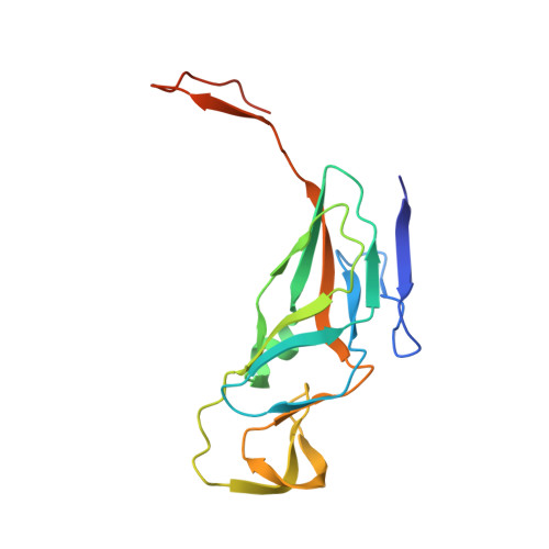 | |
UniProt | |||||
Entity Groups | |||||
| Sequence Clusters | 30% Identity50% Identity70% Identity90% Identity95% Identity100% Identity | ||||
| UniProt Group | Q8SDV3 | ||||
Sequence AnnotationsExpand | |||||
Reference Sequence | |||||
| Ligands 2 Unique | |||||
|---|---|---|---|---|---|
| ID | Chains | Name / Formula / InChI Key | 2D Diagram | 3D Interactions | |
| DUP Download:Ideal Coordinates CCD File | G [auth A] I [auth B] K [auth C] M [auth D] N [auth D] | 2'-DEOXYURIDINE 5'-ALPHA,BETA-IMIDO-TRIPHOSPHATE C9 H16 N3 O13 P3 XZLLMTSKYYYJLH-SHYZEUOFSA-N |  | ||
| MG Download:Ideal Coordinates CCD File | H [auth A] J [auth B] L [auth C] O [auth D] P [auth E] | MAGNESIUM ION Mg JLVVSXFLKOJNIY-UHFFFAOYSA-N |  | ||
| Length ( Å ) | Angle ( ˚ ) |
|---|---|
| a = 108.42 | α = 90 |
| b = 108.42 | β = 90 |
| c = 167.05 | γ = 90 |
| Software Name | Purpose |
|---|---|
| PHENIX | refinement |
| XDS | data reduction |
| XSCALE | data scaling |
| PHASER | phasing |
| Funding Organization | Location | Grant Number |
|---|---|---|
| ungarian Scientific Research Fund OTKA | Hungary | NK 84008 |
| ungarian Scientific Research Fund OTKA | Hungary | K109486 |
| Baross Program of the New Hungary Development Plan | Hungary | 3DSTRUCT, MFB-00266/2010 REG-KM-09-1-2009-005 |
| Hungarian Academy of Sciences | Hungary | TTK IF-28/201 |
| Hungarian Academy of Sciences | Hungary | MedinProt program |
| European Union | 283570 |


