4WFD

Structure of the Rrp6-Rrp47-Mtr4 interaction


Experimental Data Snapshot

  • Method: X-RAY DIFFRACTION
  • Resolution: 2.40 Å
  • R-Value Free: 0.243 
  • R-Value Work: 0.201 
  • R-Value Observed: 0.203 

wwPDB Validation   3D Report Full Report


This is version 3.0 of the entry. See complete history


Literature

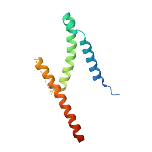
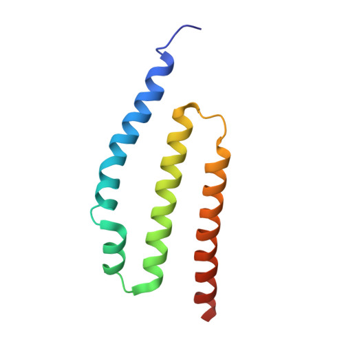
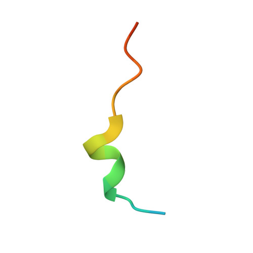
The exosome-binding factors Rrp6 and Rrp47 form a composite surface for recruiting the Mtr4 helicase.

Schuch, B.Feigenbutz, M.Makino, D.L.Falk, S.Basquin, C.Mitchell, P.Conti, E.

(2014) EMBO J 33: 2829-2846

  • DOI: https://doi.org/10.15252/embj.201488757
  • Primary Citation of Related Structures:  
    4WFC, 4WFD

  • PubMed Abstract: 

    The exosome is a conserved multi-subunit ribonuclease complex that functions in 3' end processing, turnover and surveillance of nuclear and cytoplasmic RNAs. In the yeast nucleus, the 10-subunit core complex of the exosome (Exo-10) physically and functionally interacts with the Rrp6 exoribonuclease and its associated cofactor Rrp47, the helicase Mtr4 and Mpp6. Here, we show that binding of Mtr4 to Exo-10 in vitro is dependent upon both Rrp6 and Rrp47, whereas Mpp6 binds directly and independently of other cofactors. Crystallographic analyses reveal that the N-terminal domains of Rrp6 and Rrp47 form a highly intertwined structural unit. Rrp6 and Rrp47 synergize to create a composite and conserved surface groove that binds the N-terminus of Mtr4. Mutation of conserved residues within Rrp6 and Mtr4 at the structural interface disrupts their interaction and inhibits growth of strains expressing a C-terminal GFP fusion of Mtr4. These studies provide detailed structural insight into the interaction between the Rrp6-Rrp47 complex and Mtr4, revealing an important link between Mtr4 and the core exosome.


  • Organizational Affiliation

    Structural Cell Biology Department, Max Planck Institute of Biochemistry, Martinsried, Germany.


Macromolecules
Find similar proteins by:  (by identity cutoff)  |  3D Structure
Entity ID: 1
MoleculeChains Sequence LengthOrganismDetailsImage
Exosome complex exonuclease RRP6
A, D, G
115Saccharomyces cerevisiae S288CMutation(s): 0 
Gene Names: RRP6UNC733YOR001W
EC: 3.1.13
UniProt
Find proteins for Q12149 (Saccharomyces cerevisiae (strain ATCC 204508 / S288c))
Explore Q12149 
Go to UniProtKB:  Q12149
Entity Groups  
Sequence Clusters30% Identity50% Identity70% Identity90% Identity95% Identity100% Identity
UniProt GroupQ12149
Sequence Annotations
Expand
  • Reference Sequence
Find similar proteins by:  (by identity cutoff)  |  3D Structure
Entity ID: 2
MoleculeChains Sequence LengthOrganismDetailsImage
Exosome complex protein LRP1
B, E, H
103Saccharomyces cerevisiae S288CMutation(s): 0 
Gene Names: LRP1RRP47YC1DYHR081W
UniProt
Find proteins for P38801 (Saccharomyces cerevisiae (strain ATCC 204508 / S288c))
Explore P38801 
Go to UniProtKB:  P38801
Entity Groups  
Sequence Clusters30% Identity50% Identity70% Identity90% Identity95% Identity100% Identity
UniProt GroupP38801
Sequence Annotations
Expand
  • Reference Sequence

Find similar proteins by:  Sequence   |   3D Structure  

Entity ID: 3
MoleculeChains Sequence LengthOrganismDetailsImage
ATP-dependent RNA helicase DOB1
C, F, I
20Saccharomyces cerevisiae S288CMutation(s): 0 
EC: 3.6.4.13
UniProt
Find proteins for P47047 (Saccharomyces cerevisiae (strain ATCC 204508 / S288c))
Explore P47047 
Go to UniProtKB:  P47047
Entity Groups  
Sequence Clusters30% Identity50% Identity70% Identity90% Identity95% Identity100% Identity
UniProt GroupP47047
Sequence Annotations
Expand
  • Reference Sequence
Small Molecules
Ligands 1 Unique
IDChains Name / Formula / InChI Key2D Diagram3D Interactions
YT3
Query on YT3

Download Ideal Coordinates CCD File 
AA [auth G]
BA [auth G]
CA [auth G]
DA [auth G]
EA [auth I]
AA [auth G],
BA [auth G],
CA [auth G],
DA [auth G],
EA [auth I],
J [auth A],
K [auth A],
L [auth A],
M [auth A],
N [auth A],
O [auth A],
P [auth B],
Q [auth C],
R [auth D],
S [auth D],
T [auth D],
U [auth D],
V [auth D],
W [auth E],
X [auth F],
Y [auth G],
Z [auth G]
YTTRIUM (III) ION
Y
GRTBAGCGDOYUBE-UHFFFAOYSA-N
Experimental Data & Validation

Experimental Data

  • Method: X-RAY DIFFRACTION
  • Resolution: 2.40 Å
  • R-Value Free: 0.243 
  • R-Value Work: 0.201 
  • R-Value Observed: 0.203 
  • Space Group: P 3 1 2
Unit Cell:
Length ( Å )Angle ( ˚ )
a = 142.745α = 90
b = 142.745β = 90
c = 63.45γ = 120
Software Package:
Software NamePurpose
XDSdata reduction
PHENIXrefinement
Cootmodel building
XSCALEdata scaling
PHENIXphasing

Structure Validation

View Full Validation Report


Entry History & Funding Information

Deposition Data


Funding OrganizationLocationGrant Number
European Research CouncilAdvanced Investigator Grant 294371
Marie CurieITN RNPnet
German Research FoundationGermanyDFG SFB646, SFB1035, GRK1721, FOR1680, CIPSM

Revision History  (Full details and data files)

  • Version 1.0: 2014-10-29
    Type: Initial release
  • Version 1.1: 2014-12-10
    Changes: Database references
  • Version 2.0: 2017-09-06
    Changes: Advisory, Atomic model, Author supporting evidence, Derived calculations, Refinement description
  • Version 3.0: 2024-01-10
    Changes: Atomic model, Data collection, Database references, Derived calculations, Refinement description