4WD8 | pdb_00004wd8

Crystal structure of a bacterial Bestrophin homolog from Klebsiella pneumoniae


Experimental Data Snapshot

  • Method: X-RAY DIFFRACTION
  • Resolution: 2.30 Å
  • R-Value Free: 
    0.214 (Depositor), 0.213 (DCC) 
  • R-Value Work: 
    0.196 (Depositor), 0.198 (DCC) 
  • R-Value Observed: 
    0.197 (Depositor) 

wwPDB Validation 3D Report Full Report

Validation slider image for 4WD8

This is version 1.4 of the entry. See complete history

Literature

Structure and selectivity in bestrophin ion channels.

Yang, T.Liu, Q.Kloss, B.Bruni, R.Kalathur, R.C.Guo, Y.Kloppmann, E.Rost, B.Colecraft, H.M.Hendrickson, W.A.

(2014) Science 346: 355-359

  • DOI: https://doi.org/10.1126/science.1259723
  • Primary Citation Related Structures: 
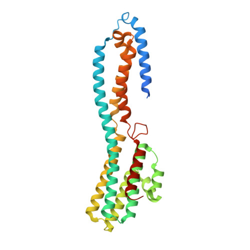
    4WD7, 4WD8

  • PubMed Abstract: 

    Human bestrophin-1 (hBest1) is a calcium-activated chloride channel from the retinal pigment epithelium, where mutations are associated with vitelliform macular degeneration, or Best disease. We describe the structure of a bacterial homolog (KpBest) of hBest1 and functional characterizations of both channels. KpBest is a pentamer that forms a five-helix transmembrane pore, closed by three rings of conserved hydrophobic residues, and has a cytoplasmic cavern with a restricted exit. From electrophysiological analysis of structure-inspired mutations in KpBest and hBest1, we find a sensitive control of ion selectivity in the bestrophins, including reversal of anion/cation selectivity, and dramatic activation by mutations at the cytoplasmic exit. A homology model of hBest1 shows the locations of disease-causing mutations and suggests possible roles in regulation.


  • Organizational Affiliation
    • Department of Biochemistry and Molecular Biophysics, Columbia University, New York, NY 10032, USA.

Macromolecule Content 

  • Total Structure Weight: 170.14 kDa 
  • Atom Count: 11,016 
  • Modeled Residue Count: 1,345 
  • Deposited Residue Count: 1,485 
  • Unique protein chains: 1

Macromolecules

Find similar proteins by:|  3D Structure
Entity ID: 1
MoleculeChains  Sequence LengthOrganismDetailsImage
Bestrophin domain protein
A, B, C, D, E
297Klebsiella pneumoniae UHKPC96Mutation(s): 0 
Gene Names: H215_4284

Small Molecules

Ligands 1 Unique
IDChains Name / Formula / InChI Key2D Diagram3D Interactions
ZN

Query on ZN


Download:Ideal Coordinates CCD File
F [auth A]
G [auth A]
H [auth A]
I [auth A]
J [auth B]
F [auth A],
G [auth A],
H [auth A],
I [auth A],
J [auth B],
K [auth B],
L [auth B],
M [auth C],
N [auth C],
O [auth C],
P [auth D],
Q [auth D],
R [auth D],
S [auth E],
T [auth E]
ZINC ION
Zn
PTFCDOFLOPIGGS-UHFFFAOYSA-N

Experimental Data & Validation

Experimental Data

  • Method: X-RAY DIFFRACTION
  • Resolution: 2.30 Å
  • R-Value Free:  0.214 (Depositor), 0.213 (DCC) 
  • R-Value Work:  0.196 (Depositor), 0.198 (DCC) 
  • R-Value Observed: 0.197 (Depositor) 
Space Group: P 21 21 21
Unit Cell:
Length ( Å )Angle ( ˚ )
a = 114.079α = 90
b = 160.034β = 90
c = 161.881γ = 90
Software Package:
Software NamePurpose
PHENIXrefinement

Structure Validation

View Full Validation Report


Entry History 

& Funding Information

Deposition Data


Funding OrganizationLocationGrant Number
National Institutes of Health/National Institute of General Medical Sciences (NIH/NIGMS)United StatesGM095315
National Institutes of Health/National Institute of General Medical Sciences (NIH/NIGMS)United StatesGM107462

Revision History  (Full details and data files)

  • Version 1.0: 2014-10-01
    Type: Initial release
  • Version 1.1: 2014-10-29
    Changes: Database references
  • Version 1.2: 2017-09-06
    Changes: Author supporting evidence, Database references, Derived calculations, Other, Source and taxonomy, Structure summary
  • Version 1.3: 2019-12-25
    Changes: Author supporting evidence
  • Version 1.4: 2023-12-27
    Changes: Data collection, Database references, Derived calculations, Refinement description