4W1Y

Crystal structure of Escherichia coli Tryptophanase in 'semi-holo' form


Experimental Data Snapshot

  • Method: X-RAY DIFFRACTION
  • Resolution: 3.20 Å
  • R-Value Free: 0.290 
  • R-Value Work: 0.214 
  • R-Value Observed: 0.218 

wwPDB Validation   3D Report Full Report


This is version 2.1 of the entry. See complete history


Literature

Structures of Escherichia coli tryptophanase in holo and `semi-holo' forms.

Kogan, A.Raznov, L.Gdalevsky, G.Y.Cohen-Luria, R.Almog, O.Parola, A.H.Goldgur, Y.

(2015) Acta Crystallogr F Struct Biol Commun 71: 286-290

  • DOI: https://doi.org/10.1107/S2053230X15000850
  • Primary Citation of Related Structures:  
    4W1Y, 4W4H

  • PubMed Abstract: 

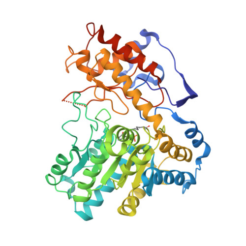
    Two crystal forms of Escherichia coli tryptophanase (tryptophan indole-lyase, Trpase) were obtained under the same crystallization conditions. Both forms belonged to the same space group P43212 but had slightly different unit-cell parameters. The holo crystal form, with pyridoxal phosphate (PLP) bound to Lys270 of both polypeptide chains in the asymmetric unit, diffracted to 2.9 Å resolution. The second crystal form diffracted to 3.2 Å resolution. Of the two subunits in the asymmetric unit, one was found in the holo form, while the other appeared to be in the apo form in a wide-open conformation with two sulfate ions bound in the vicinity of the active site. The conformation of all holo subunits is the same in both crystal forms. The structures suggest that Trpase is flexible in the apo form. Its conformation partially closes upon binding of PLP. The closed conformation might correspond to the enzyme in its active state with both cofactor and substrate bound in a similar way as in tyrosine phenol-lyase.


  • Organizational Affiliation

    Department of Chemistry, Ben Gurion University of the Negev, Beer Sheva 84105, Israel.


Macromolecules
Find similar proteins by:  (by identity cutoff)  |  3D Structure
Entity ID: 1
MoleculeChains Sequence LengthOrganismDetailsImage
Tryptophanase467Escherichia coli K-12Mutation(s): 0 
Gene Names: tnaAindb3708JW3686
EC: 4.1.99.1
UniProt
Find proteins for P0A853 (Escherichia coli (strain K12))
Explore P0A853 
Go to UniProtKB:  P0A853
Entity Groups  
Sequence Clusters30% Identity50% Identity70% Identity90% Identity95% Identity100% Identity
UniProt GroupP0A853
Sequence Annotations
Expand
  • Reference Sequence
Find similar proteins by:  (by identity cutoff)  |  3D Structure
Entity ID: 2
MoleculeChains Sequence LengthOrganismDetailsImage
Tryptophanase467Escherichia coli K-12Mutation(s): 0 
Gene Names: tnaAindb3708JW3686
EC: 4.1.99.1
UniProt
Find proteins for P0A853 (Escherichia coli (strain K12))
Explore P0A853 
Go to UniProtKB:  P0A853
Entity Groups  
Sequence Clusters30% Identity50% Identity70% Identity90% Identity95% Identity100% Identity
UniProt GroupP0A853
Sequence Annotations
Expand
  • Reference Sequence
Small Molecules
Modified Residues  1 Unique
IDChains TypeFormula2D DiagramParent
LLP
Query on LLP
A
L-PEPTIDE LINKINGC14 H22 N3 O7 PLYS
Experimental Data & Validation

Experimental Data

  • Method: X-RAY DIFFRACTION
  • Resolution: 3.20 Å
  • R-Value Free: 0.290 
  • R-Value Work: 0.214 
  • R-Value Observed: 0.218 
  • Space Group: P 43 21 2
Unit Cell:
Length ( Å )Angle ( ˚ )
a = 109.971α = 90
b = 109.971β = 90
c = 238.403γ = 90
Software Package:
Software NamePurpose
REFMACrefinement

Structure Validation

View Full Validation Report


Entry History 

Deposition Data

  • Released Date: 2014-12-17 
  • Deposition Author(s): Goldgur, Y.

Revision History  (Full details and data files)

  • Version 1.0: 2014-12-17
    Type: Initial release
  • Version 1.1: 2015-03-11
    Changes: Database references
  • Version 1.2: 2015-03-25
    Changes: Database references
  • Version 2.0: 2017-09-27
    Changes: Data collection, Derived calculations, Non-polymer description, Source and taxonomy, Structure summary
  • Version 2.1: 2023-12-27
    Changes: Data collection, Database references