4V07 | pdb_00004v07

Dimeric pseudorabies virus protease pUL26N at 2.1 A resolution


Experimental Data Snapshot

  • Method: X-RAY DIFFRACTION
  • Resolution: 2.10 Å
  • R-Value Free: 
    0.237 (Depositor), 0.242 (DCC) 
  • R-Value Work: 
    0.178 (Depositor), 0.186 (DCC) 
  • R-Value Observed: 
    0.181 (Depositor) 

Starting Model: experimental
View more details

wwPDB Validation 3D Report Full Report

Validation slider image for 4V07

This is version 1.2 of the entry. See complete history

Literature

Dimerization-Induced Allosteric Changes of the Oxyanion-Hole Loop Activate the Pseudorabies Virus Assemblin Pul26N, a Herpesvirus Serine Protease

Zuehlsdorf, M.Werten, S.Klupp, B.G.Palm, G.J.Mettenleiter, T.Hinrichs, W.

(2015) PLoS Pathog 11: 5045

  • DOI: https://doi.org/10.1371/journal.ppat.1005045
  • Primary Citation Related Structures: 
    4CX8, 4V07, 4V08, 4V0T

  • PubMed Abstract: 

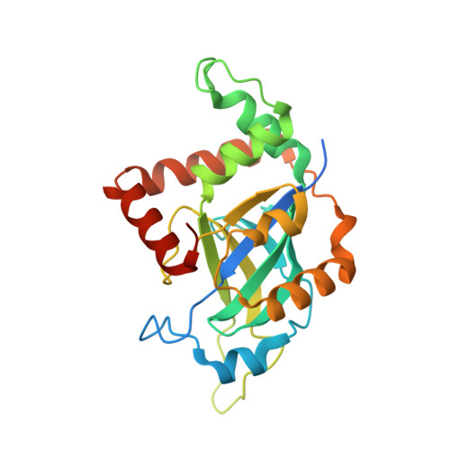
    Herpesviruses encode a characteristic serine protease with a unique fold and an active site that comprises the unusual triad Ser-His-His. The protease is essential for viral replication and as such constitutes a promising drug target. In solution, a dynamic equilibrium exists between an inactive monomeric and an active dimeric form of the enzyme, which is believed to play a key regulatory role in the orchestration of proteolysis and capsid assembly. Currently available crystal structures of herpesvirus proteases correspond either to the dimeric state or to complexes with peptide mimetics that alter the dimerization interface. In contrast, the structure of the native monomeric state has remained elusive. Here, we present the three-dimensional structures of native monomeric, active dimeric, and diisopropyl fluorophosphate-inhibited dimeric protease derived from pseudorabies virus, an alphaherpesvirus of swine. These structures, solved by X-ray crystallography to respective resolutions of 2.05, 2.10 and 2.03 Å, allow a direct comparison of the main conformational states of the protease. In the dimeric form, a functional oxyanion hole is formed by a loop of 10 amino-acid residues encompassing two consecutive arginine residues (Arg136 and Arg137); both are strictly conserved throughout the herpesviruses. In the monomeric form, the top of the loop is shifted by approximately 11 Å, resulting in a complete disruption of the oxyanion hole and loss of activity. The dimerization-induced allosteric changes described here form the physical basis for the concentration-dependent activation of the protease, which is essential for proper virus replication. Small-angle X-ray scattering experiments confirmed a concentration-dependent equilibrium of monomeric and dimeric protease in solution.


  • Organizational Affiliation
    • Institute of Biochemistry, University of Greifswald, Greifswald, Germany.

Macromolecule Content 

  • Total Structure Weight: 53.56 kDa 
  • Atom Count: 3,477 
  • Modeled Residue Count: 442 
  • Deposited Residue Count: 488 
  • Unique protein chains: 1

Macromolecules

Find similar proteins by:|  3D Structure
Entity ID: 1
MoleculeChains  Sequence LengthOrganismDetailsImage
UL26
A, B
244Suid alphaherpesvirus 1Mutation(s): 0 
EC: 3.4.21.97
UniProt
Find proteins for Q83417 (Suid herpesvirus 1 (strain Kaplan))
Explore Q83417 
Go to UniProtKB:  Q83417
Entity Groups
Sequence Clusters30% Identity50% Identity70% Identity90% Identity95% Identity100% Identity
UniProt GroupQ83417
Sequence Annotations
Expand
Reference Sequence

Small Molecules

Ligands 2 Unique
IDChains Name / Formula / InChI Key2D Diagram3D Interactions
CL

Query on CL


Download:Ideal Coordinates CCD File
C [auth A]
D [auth A]
E [auth A]
F [auth A]
H [auth B]
C [auth A],
D [auth A],
E [auth A],
F [auth A],
H [auth B],
I [auth B],
J [auth B]
CHLORIDE ION
Cl
VEXZGXHMUGYJMC-UHFFFAOYSA-M
MG

Query on MG


Download:Ideal Coordinates CCD File
G [auth A]MAGNESIUM ION
Mg
JLVVSXFLKOJNIY-UHFFFAOYSA-N

Experimental Data & Validation

Experimental Data

  • Method: X-RAY DIFFRACTION
  • Resolution: 2.10 Å
  • R-Value Free:  0.237 (Depositor), 0.242 (DCC) 
  • R-Value Work:  0.178 (Depositor), 0.186 (DCC) 
  • R-Value Observed: 0.181 (Depositor) 
Space Group: P 21 21 21
Unit Cell:
Length ( Å )Angle ( ˚ )
a = 51.375α = 90
b = 75.986β = 90
c = 110.758γ = 90
Software Package:
Software NamePurpose
REFMACrefinement
XDSdata reduction
Aimlessdata scaling
PHASERphasing

Structure Validation

View Full Validation Report


Entry History 

Deposition Data

Revision History  (Full details and data files)

  • Version 1.0: 2015-07-15
    Type: Initial release
  • Version 1.1: 2015-07-22
    Changes: Database references
  • Version 1.2: 2024-01-10
    Changes: Data collection, Database references, Derived calculations, Other, Refinement description