Structural Mechanism of Glutamate Receptor Activation and Desensitization
Meyerson, J.R., Kumar, J., Chittori, S., Rao, P., Pierson, J., Bartesaghi, A., Mayer, M.L., Subramaniam, S.(2014) Nature 514: 328
- PubMed: 25119039 Search on PubMedSearch on PubMed Central
- DOI: https://doi.org/10.1038/nature13603
- Primary Citation Related Structures:
4UQ6, 4UQJ, 4UQK, 4UQQ - PubMed Abstract:
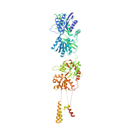
Ionotropic glutamate receptors are ligand-gated ion channels that mediate excitatory synaptic transmission in the vertebrate brain. To gain a better understanding of how structural changes gate ion flux across the membrane, we trapped rat AMPA (α-amino-3-hydroxy-5-methyl-4-isoxazole propionic acid) and kainate receptor subtypes in their major functional states and analysed the resulting structures using cryo-electron microscopy. We show that transition to the active state involves a 'corkscrew' motion of the receptor assembly, driven by closure of the ligand-binding domain. Desensitization is accompanied by disruption of the amino-terminal domain tetramer in AMPA, but not kainate, receptors with a two-fold to four-fold symmetry transition in the ligand-binding domains in both subtypes. The 7.6 Å structure of a desensitized kainate receptor shows how these changes accommodate channel closing. These findings integrate previous physiological, biochemical and structural analyses of glutamate receptors and provide a molecular explanation for key steps in receptor gating.
- Laboratory of Cell Biology, Center for Cancer Research, NCI, NIH, Bethesda, Maryland 20892, USA.
Organizational Affiliation: