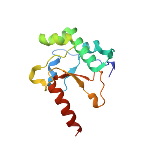
Structures of the Ultra-High Affinity Protein-Protein Complexes of Pyocins S2 and Ap41 and Their Cognate Immunity Proteins from Pseudomonas Aeruginosa
Joshi, A., Grinter, R., Josts, I., Chen, S., Wojdyla, J.A., Lowe, E.D., Kaminska, R., Sharp, C., Mccaughey, L., Roszak, A.W., Cogdell, R.J., Byron, O., Walker, D., Kleanthous, C.(2015) J Mol Biology 427: 2852
- PubMed: 26215615
- DOI: https://doi.org/10.1016/j.jmb.2015.07.014
- Primary Citation of Related Structures:
4UHP, 4UHQ - PubMed Abstract:
How ultra-high-affinity protein-protein interactions retain high specificity is still poorly understood. The interaction between colicin DNase domains and their inhibitory immunity (Im) proteins is an ultra-high-affinity interaction that is essential for the neutralisation of endogenous DNase catalytic activity and for protection against exogenous DNase bacteriocins. The colicin DNase-Im interaction is a model system for the study of high-affinity protein-protein interactions. However, despite the fact that closely related colicin-like bacteriocins are widely produced by Gram-negative bacteria, this interaction has only been studied using colicins from Escherichia coli. In this work, we present the first crystal structures of two pyocin DNase-Im complexes from Pseudomonas aeruginosa, pyocin S2 DNase-ImS2 and pyocin AP41 DNase-ImAP41. These structures represent divergent DNase-Im subfamilies and are important in extending our understanding of protein-protein interactions for this important class of high-affinity protein complex. A key finding of this work is that mutations within the immunity protein binding energy hotspot, helix III, are tolerated by complementary substitutions at the DNase-Immunity protein binding interface. Im helix III is strictly conserved in colicins where an Asp forms polar interactions with the DNase backbone. ImAP41 contains an Asp-to-Gly substitution in helix III and our structures show the role of a co-evolved substitution where Pro in DNase loop 4 occupies the volume vacated and removes the unfulfilled hydrogen bond. We observe the co-evolved mutations in other DNase-Immunity pairs that appear to underpin the split of this family into two distinct groups.
- Department of Biochemistry, University of Oxford, South Parks Road, Oxford OX1 3QU, UK.
Organizational Affiliation:


