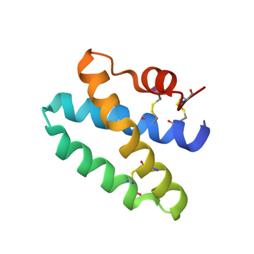
4UEX

Structure of human Saposin A at lysosomal pH


Experimental Data Snapshot

  • Method: X-RAY DIFFRACTION
  • Resolution: 1.80 Å
  • R-Value Free: 0.210 
  • R-Value Work: 0.185 
  • R-Value Observed: 0.186 

wwPDB Validation   3D Report Full Report


This is version 1.2 of the entry. See complete history


Literature

Structure of Human Saposin a at Lysosomal Ph.

Hill, C.H.Read, R.J.Deane, J.E.

(2015) Acta Crystallogr D Biol Crystallogr 71: 895

  • DOI: https://doi.org/10.1107/S2053230X15008584
  • Primary Citation of Related Structures:  
    4UEX

  • PubMed Abstract: 

    The saposins are essential cofactors for the normal lysosomal degradation of complex glycosphingolipids by acid hydrolase enzymes; defects in either saposin or hydrolase function lead to severe metabolic diseases. Saposin A (SapA) activates the enzyme β-galactocerebrosidase (GALC), which catalyzes the breakdown of β-D-galactocerebroside, the principal lipid component of myelin. SapA is known to bind lipids and detergents in a pH-dependent manner; this is accompanied by a striking transition from a `closed' to an `open' conformation. However, previous structures were determined at non-lysosomal pH. This work describes a 1.8 Å resolution X-ray crystal structure determined at the physiologically relevant lysosomal pH 4.8. In the absence of lipid or detergent at pH 4.8, SapA is observeed to adopt a conformation closely resembling the previously determined `closed' conformation, showing that pH alone is not sufficient for the transition to the `open' conformation. Structural alignments reveal small conformational changes, highlighting regions of flexibility.


  • Organizational Affiliation

    Department of Haematology, Cambridge Institute for Medical Research, University of Cambridge, Wellcome Trust/MRC Building, Cambridge Biomedical Campus, Hills Road, Cambridge CB2 0XY, England.


Macromolecules
Find similar proteins by:  (by identity cutoff)  |  3D Structure
Entity ID: 1
MoleculeChains Sequence LengthOrganismDetailsImage
PROSAPOSIN
A, B
85Homo sapiensMutation(s): 0 
UniProt & NIH Common Fund Data Resources
Find proteins for P07602 (Homo sapiens)
Explore P07602 
Go to UniProtKB:  P07602
PHAROS:  P07602
GTEx:  ENSG00000197746 
Entity Groups  
Sequence Clusters30% Identity50% Identity70% Identity90% Identity95% Identity100% Identity
UniProt GroupP07602
Sequence Annotations
Expand
  • Reference Sequence
Experimental Data & Validation

Experimental Data

  • Method: X-RAY DIFFRACTION
  • Resolution: 1.80 Å
  • R-Value Free: 0.210 
  • R-Value Work: 0.185 
  • R-Value Observed: 0.186 
  • Space Group: P 1 21 1
Unit Cell:
Length ( Å )Angle ( ˚ )
a = 34.17α = 90
b = 58.69β = 93.1
c = 35.16γ = 90
Software Package:
Software NamePurpose
PHENIXrefinement
XDSdata reduction
Aimlessdata scaling
PHASERphasing

Structure Validation

View Full Validation Report


Entry History 

Deposition Data

Revision History  (Full details and data files)

  • Version 1.0: 2015-07-15
    Type: Initial release
  • Version 1.1: 2019-05-29
    Changes: Data collection, Experimental preparation, Other
  • Version 1.2: 2023-12-20
    Changes: Data collection, Database references, Other, Refinement description