4RLE | pdb_00004rle

Crystal structure of the c-di-AMP binding PII-like protein DarA


Experimental Data Snapshot

  • Method: X-RAY DIFFRACTION
  • Resolution: 1.30 Å
  • R-Value Free: 
    0.154 (Depositor), 0.156 (DCC) 
  • R-Value Work: 
    0.130 (Depositor), 0.134 (DCC) 
  • R-Value Observed: 
    0.131 (Depositor) 

Starting Model: experimental
View more details

wwPDB Validation 3D Report Full Report

Validation slider image for 4RLE

Ligand Structure Quality Assessment 


This is version 1.4 of the entry. See complete history

Literature

Identification, Characterization, and Structure Analysis of the Cyclic di-AMP-binding PII-like Signal Transduction Protein DarA.

Gundlach, J.Dickmanns, A.Schroder-Tittmann, K.Neumann, P.Kaesler, J.Kampf, J.Herzberg, C.Hammer, E.Schwede, F.Kaever, V.Tittmann, K.Stulke, J.Ficner, R.

(2015) J Biological Chem 290: 3069-3080

  • DOI: https://doi.org/10.1074/jbc.M114.619619
  • Primary Citation Related Structures: 
    4RLE

  • PubMed Abstract: 

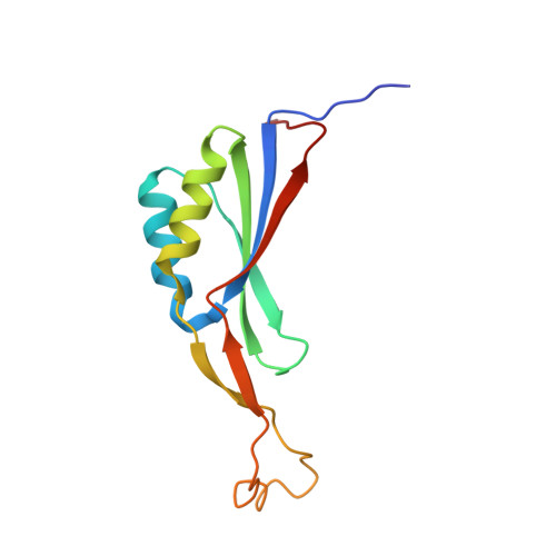
    The cyclic dimeric AMP nucleotide c-di-AMP is an essential second messenger in Bacillus subtilis. We have identified the protein DarA as one of the prominent c-di-AMP receptors in B. subtilis. Crystal structure analysis shows that DarA is highly homologous to PII signal transducer proteins. In contrast to PII proteins, the functionally important B- and T-loops are swapped with respect to their size. DarA is a homotrimer that binds three molecules of c-di-AMP, each in a pocket located between two subunits. We demonstrate that DarA is capable to bind c-di-AMP and with lower affinity cyclic GMP-AMP (3'3'-cGAMP) but not c-di-GMP or 2'3'-cGAMP. Consistently the crystal structure shows that within the ligand-binding pocket only one adenine is highly specifically recognized, whereas the pocket for the other adenine appears to be promiscuous. Comparison with a homologous ligand-free DarA structure reveals that c-di-AMP binding is accompanied by conformational changes of both the fold and the position of the B-loop in DarA.


  • Organizational Affiliation
    • From the Departments of General Microbiology.

Macromolecule Content 

  • Total Structure Weight: 13.67 kDa 
  • Atom Count: 1,204 
  • Modeled Residue Count: 116 
  • Deposited Residue Count: 117 
  • Unique protein chains: 1

Macromolecules

Find similar proteins by:|  3D Structure
Entity ID: 1
MoleculeChains  Sequence LengthOrganismDetailsImage
Uncharacterized protein YaaQ117Bacillus subtilis subsp. subtilis str. 168Mutation(s): 0 
Gene Names: BSU00290yaaQ
UniProt
Find proteins for P37538 (Bacillus subtilis (strain 168))
Explore P37538 
Go to UniProtKB:  P37538
Entity Groups
Sequence Clusters30% Identity50% Identity70% Identity90% Identity95% Identity100% Identity
UniProt GroupP37538
Sequence Annotations
Expand
Reference Sequence

Small Molecules

Ligands 2 Unique
IDChains Name / Formula / InChI Key2D Diagram3D Interactions
2BA

Query on 2BA


Download:Ideal Coordinates CCD File
B [auth A](2R,3R,3aS,5R,7aR,9R,10R,10aS,12R,14aR)-2,9-bis(6-amino-9H-purin-9-yl)octahydro-2H,7H-difuro[3,2-d:3',2'-j][1,3,7,9,2,8 ]tetraoxadiphosphacyclododecine-3,5,10,12-tetrol 5,12-dioxide
C20 H24 N10 O12 P2
PDXMFTWFFKBFIN-XPWFQUROSA-N
NI

Query on NI


Download:Ideal Coordinates CCD File
C [auth A]NICKEL (II) ION
Ni
VEQPNABPJHWNSG-UHFFFAOYSA-N

Experimental Data & Validation

Experimental Data

  • Method: X-RAY DIFFRACTION
  • Resolution: 1.30 Å
  • R-Value Free:  0.154 (Depositor), 0.156 (DCC) 
  • R-Value Work:  0.130 (Depositor), 0.134 (DCC) 
  • R-Value Observed: 0.131 (Depositor) 
Space Group: P 63
Unit Cell:
Length ( Å )Angle ( ˚ )
a = 56.521α = 90
b = 56.521β = 90
c = 58.201γ = 120
Software Package:
Software NamePurpose
XSCALEdata scaling
PHENIXrefinement
PDB_EXTRACTdata extraction
DNAdata collection
XDSdata reduction
PHASERphasing

Structure Validation

View Full Validation Report


Ligand Structure Quality Assessment 


Entry History 

Deposition Data

Revision History  (Full details and data files)

  • Version 1.0: 2014-12-03
    Type: Initial release
  • Version 1.1: 2014-12-17
    Changes: Database references
  • Version 1.2: 2015-02-11
    Changes: Database references
  • Version 1.3: 2019-09-04
    Changes: Data collection
  • Version 1.4: 2023-09-20
    Changes: Data collection, Database references, Derived calculations, Refinement description, Structure summary