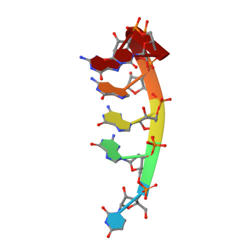
Structural Variations and Solvent Structure of r(UGGGGU) Quadruplexes Stabilized by Sr(2+) Ions.
Fyfe, A.C., Dunten, P.W., Martick, M.M., Scott, W.G.(2015) J Mol Biology 427: 2205-2219
- PubMed: 25861762
- DOI: https://doi.org/10.1016/j.jmb.2015.03.022
- Primary Citation of Related Structures:
4RJ1, 4RKV, 4RNE - PubMed Abstract:
Guanine-rich sequences can, under appropriate conditions, adopt a distinctive, four-stranded, helical fold known as a G-quadruplex. Interest in quadruplex folds has grown in recent years as evidence of their biological relevance has accumulated from both sequence analysis and function-specific assays. The folds are unusually stable and their formation appears to require close management to maintain cell health; regulatory failure correlates with genomic instability and a number of cancer phenotypes. Biologically relevant quadruplex folds are anticipated to form transiently in mRNA and in single-stranded, unwound DNA. To elucidate factors, including bound solvent, that contribute to the stability of RNA quadruplexes, we examine, by X-ray crystallography and small-angle X-ray scattering, the structure of a previously reported tetramolecular quadruplex, UGGGGU stabilized by Sr(2+) ions. Crystal forms of the octameric assembly formed by this sequence exhibit unusually strong diffraction and anomalous signal enabling the construction of reliable models to a resolution of 0.88Å. The solvent structure confirms hydration patterns reported for other nucleic acid helical conformations and provides support for the greater stability of RNA quadruplexes relative to DNA. Novel features detected in the octameric RNA assembly include a new crystal form, evidence of multiple conformations and structural variations in the 3' U tetrad, including one that leads to the formation of a hydrated internal cavity.
- Department of Chemistry and Biochemistry, University of California, Santa Cruz, CA 95064, USA.
Organizational Affiliation:


