4RH7

Crystal structure of human cytoplasmic dynein 2 motor domain in complex with ADP.Vi


Experimental Data Snapshot

  • Method: X-RAY DIFFRACTION
  • Resolution: 3.41 Å
  • R-Value Free: 0.285 
  • R-Value Work: 0.237 
  • R-Value Observed: 0.240 

wwPDB Validation   3D Report Full Report


Ligand Structure Quality Assessment 


This is version 1.4 of the entry. See complete history


Literature

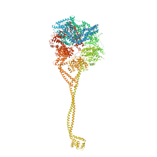
Structure of human cytoplasmic dynein-2 primed for its power stroke.

Schmidt, H.Zalyte, R.Urnavicius, L.Carter, A.P.

(2015) Nature 518: 435-438

  • DOI: https://doi.org/10.1038/nature14023
  • Primary Citation of Related Structures:  
    4RH7

  • PubMed Abstract: 

    Members of the dynein family, consisting of cytoplasmic and axonemal isoforms, are motors that move towards the minus ends of microtubules. Cytoplasmic dynein-1 (dynein-1) plays roles in mitosis and cellular cargo transport, and is implicated in viral infections and neurodegenerative diseases. Cytoplasmic dynein-2 (dynein-2) performs intraflagellar transport and is associated with human skeletal ciliopathies. Dyneins share a conserved motor domain that couples cycles of ATP hydrolysis with conformational changes to produce movement. Here we present the crystal structure of the human cytoplasmic dynein-2 motor bound to the ATP-hydrolysis transition state analogue ADP.vanadate. The structure reveals a closure of the motor's ring of six AAA+ domains (ATPases associated with various cellular activites: AAA1-AAA6). This induces a steric clash with the linker, the key element for the generation of movement, driving it into a conformation that is primed to produce force. Ring closure also changes the interface between the stalk and buttress coiled-coil extensions of the motor domain. This drives helix sliding in the stalk which causes the microtubule binding domain at its tip to release from the microtubule. Our structure answers the key questions of how ATP hydrolysis leads to linker remodelling and microtubule affinity regulation.


  • Organizational Affiliation

    Medical Research Council Laboratory of Molecular Biology, Division of Structural Studies, Francis Crick Avenue, Cambridge, CB2 0QH, United Kingdom.


Macromolecules
Find similar proteins by:  (by identity cutoff)  |  3D Structure
Entity ID: 1
MoleculeChains Sequence LengthOrganismDetailsImage
Green fluorescent protein/Cytoplasmic dynein 2 heavy chain 13,450synthetic constructHomo sapiens
This entity is chimeric
Mutation(s): 3 
UniProt & NIH Common Fund Data Resources
Find proteins for Q8NCM8 (Homo sapiens)
Explore Q8NCM8 
Go to UniProtKB:  Q8NCM8
PHAROS:  Q8NCM8
GTEx:  ENSG00000187240 
Entity Groups  
Sequence Clusters30% Identity50% Identity70% Identity90% Identity95% Identity100% Identity
UniProt GroupQ8NCM8
Sequence Annotations
Expand
  • Reference Sequence
Small Molecules
Experimental Data & Validation

Experimental Data

  • Method: X-RAY DIFFRACTION
  • Resolution: 3.41 Å
  • R-Value Free: 0.285 
  • R-Value Work: 0.237 
  • R-Value Observed: 0.240 
  • Space Group: C 2 2 21
Unit Cell:
Length ( Å )Angle ( ˚ )
a = 136.03α = 90
b = 487.15β = 90
c = 276.46γ = 90
Software Package:
Software NamePurpose
SHARPphasing
REFMACrefinement
MOSFLMdata reduction
SCALAdata scaling

Structure Validation

View Full Validation Report


Ligand Structure Quality Assessment 


Entry History 

Deposition Data

Revision History  (Full details and data files)

  • Version 1.0: 2014-12-10
    Type: Initial release
  • Version 1.1: 2014-12-24
    Changes: Database references
  • Version 1.2: 2015-04-08
    Changes: Database references
  • Version 1.3: 2017-08-02
    Changes: Refinement description, Source and taxonomy
  • Version 1.4: 2024-02-28
    Changes: Data collection, Database references, Derived calculations