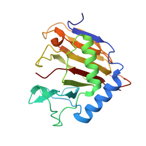
4RFR

Complex structure of AlkB/rhein


Experimental Data Snapshot

  • Method: X-RAY DIFFRACTION
  • Resolution: 1.50 Å
  • R-Value Free: 0.150 
  • R-Value Work: 0.118 
  • R-Value Observed: 0.119 

wwPDB Validation   3D Report Full Report


Ligand Structure Quality Assessment 


This is version 1.2 of the entry. See complete history


Literature

Rhein Inhibits AlkB Repair Enzymes and Sensitizes Cells to Methylated DNA Damage.

Li, Q.Huang, Y.Liu, X.Gan, J.Chen, H.Yang, C.G.

(2016) J Biol Chem 291: 11083-11093

  • DOI: https://doi.org/10.1074/jbc.M115.711895
  • Primary Citation of Related Structures:  
    4RFR

  • PubMed Abstract: 

    The AlkB repair enzymes, including Escherichia coli AlkB and two human homologues, ALKBH2 and ALKBH3, are iron(II)- and 2-oxoglutarate-dependent dioxygenases that efficiently repair N(1)-methyladenine and N(3)-methylcytosine methylated DNA damages. The development of small molecule inhibitors of these enzymes has seen less success. Here we have characterized a previously discovered natural product rhein and tested its ability to inhibit AlkB repair enzymes in vitro and to sensitize cells to methyl methane sulfonate that mainly produces N(1)-methyladenine and N(3)-methylcytosine lesions. Our investigation of the mechanism of rhein inhibition reveals that rhein binds to AlkB repair enzymes in vitro and promotes thermal stability in vivo In addition, we have determined a new structural complex of rhein bound to AlkB, which shows that rhein binds to a different part of the active site in AlkB than it binds to in fat mass and obesity-associated protein (FTO). With the support of these observations, we put forth the hypothesis that AlkB repair enzymes would be effective pharmacological targets for cancer treatment.


  • Organizational Affiliation

    From the Laboratory of Chemical Biology, State Key Laboratory of Drug Research, Shanghai Institute of Materia Medica, Chinese Academy of Sciences, Shanghai 201203, China.


Macromolecules
Find similar proteins by:  (by identity cutoff)  |  3D Structure
Entity ID: 1
MoleculeChains Sequence LengthOrganismDetailsImage
Alpha-ketoglutarate-dependent dioxygenase AlkBA [auth B]203Escherichia coliMutation(s): 0 
Gene Names: alkBaidD
EC: 1.14.11.33
UniProt
Find proteins for P05050 (Escherichia coli (strain K12))
Explore P05050 
Go to UniProtKB:  P05050
Entity Groups  
Sequence Clusters30% Identity50% Identity70% Identity90% Identity95% Identity100% Identity
UniProt GroupP05050
Sequence Annotations
Expand
  • Reference Sequence
Small Molecules
Ligands 2 Unique
IDChains Name / Formula / InChI Key2D Diagram3D Interactions
RHN
Query on RHN

Download Ideal Coordinates CCD File 
B
4,5-dihydroxy-9,10-dioxo-9,10-dihydroanthracene-2-carboxylic acid
C15 H8 O6
FCDLCPWAQCPTKC-UHFFFAOYSA-N
MN
Query on MN

Download Ideal Coordinates CCD File 
C [auth B]MANGANESE (II) ION
Mn
WAEMQWOKJMHJLA-UHFFFAOYSA-N
Binding Affinity Annotations 
IDSourceBinding Affinity
RHN Binding MOAD:  4RFR Kd: 290 (nM) from 1 assay(s)
Experimental Data & Validation

Experimental Data

  • Method: X-RAY DIFFRACTION
  • Resolution: 1.50 Å
  • R-Value Free: 0.150 
  • R-Value Work: 0.118 
  • R-Value Observed: 0.119 
  • Space Group: P 1
Unit Cell:
Length ( Å )Angle ( ˚ )
a = 36.95α = 77.37
b = 39.2β = 74.67
c = 40.521γ = 65.47
Software Package:
Software NamePurpose
HKL-2000data collection
PHASESphasing
REFMACrefinement
HKL-2000data reduction
HKL-2000data scaling

Structure Validation

View Full Validation Report


Ligand Structure Quality Assessment 


Entry History 

Deposition Data

Revision History  (Full details and data files)

  • Version 1.0: 2016-04-06
    Type: Initial release
  • Version 1.1: 2016-09-28
    Changes: Database references
  • Version 1.2: 2024-02-28
    Changes: Data collection, Database references, Derived calculations