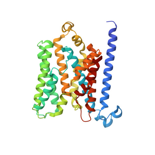
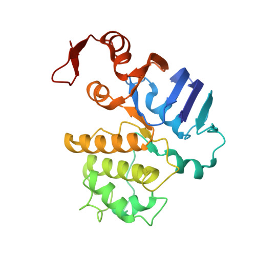
4R9U

Structure of vitamin B12 transporter BtuCD in a nucleotide-bound outward facing state


Experimental Data Snapshot

  • Method: X-RAY DIFFRACTION
  • Resolution: 2.79 Å
  • R-Value Free: 0.246 
  • R-Value Work: 0.217 
  • R-Value Observed: 0.218 

Starting Model: experimental
View more details

wwPDB Validation   3D Report Full Report


Ligand Structure Quality Assessment 


This is version 1.3 of the entry. See complete history


Literature

Structure of AMP-PNP-bound BtuCD and mechanism of ATP-powered vitamin B12 transport by BtuCD-F.

Korkhov, V.M.Mireku, S.A.Veprintsev, D.B.Locher, K.P.

(2014) Nat Struct Mol Biol 21: 1097-1099

  • DOI: https://doi.org/10.1038/nsmb.2918
  • Primary Citation of Related Structures:  
    4R9U

  • PubMed Abstract: 

    The reaction mechanism of BtuCD-F-catalyzed vitamin B12 transport into Escherichia coli is currently unclear. Here we present the structure of the last missing state in the form of AMP-PNP-bound BtuCD, trapped by a disulfide cross-link. Our structural and biochemical data allow a consistent mechanism to be formulated, thus rationalizing the roles of substrate, ATP and substrate-binding protein.


  • Organizational Affiliation

    1] Institute of Molecular Biology and Biophysics, Eidgenössische Technische Hochschule Zurich, Zurich, Switzerland. [2] Laboratory of Biomolecular Research, Paul Scherrer Institute, Villigen, Switzerland.


Macromolecules
Find similar proteins by:  (by identity cutoff)  |  3D Structure
Entity ID: 1
MoleculeChains Sequence LengthOrganismDetailsImage
Vitamin B12 import system permease protein BtuC
A, B
333Escherichia coli K-12Mutation(s): 7 
Gene Names: btuCb1711JW1701
EC: 3.6.3.33
Membrane Entity: Yes 
UniProt
Find proteins for P06609 (Escherichia coli (strain K12))
Explore P06609 
Go to UniProtKB:  P06609
Entity Groups  
Sequence Clusters30% Identity50% Identity70% Identity90% Identity95% Identity100% Identity
UniProt GroupP06609
Sequence Annotations
Expand
  • Reference Sequence
Find similar proteins by:  (by identity cutoff)  |  3D Structure
Entity ID: 2
MoleculeChains Sequence LengthOrganismDetailsImage
Vitamin B12 import ATP-binding protein BtuD
C, D
249Escherichia coli K-12Mutation(s): 3 
Gene Names: btuDb1709JW1699
EC: 3.6.3.33
Membrane Entity: Yes 
UniProt
Find proteins for P06611 (Escherichia coli (strain K12))
Explore P06611 
Go to UniProtKB:  P06611
Entity Groups  
Sequence Clusters30% Identity50% Identity70% Identity90% Identity95% Identity100% Identity
UniProt GroupP06611
Sequence Annotations
Expand
  • Reference Sequence
Small Molecules
Ligands 3 Unique
IDChains Name / Formula / InChI Key2D Diagram3D Interactions
ANP
Query on ANP

Download Ideal Coordinates CCD File 
I [auth C],
K [auth D]
PHOSPHOAMINOPHOSPHONIC ACID-ADENYLATE ESTER
C10 H17 N6 O12 P3
PVKSNHVPLWYQGJ-KQYNXXCUSA-N
LDA
Query on LDA

Download Ideal Coordinates CCD File 
E [auth A],
F [auth A],
G [auth B],
H [auth B]
LAURYL DIMETHYLAMINE-N-OXIDE
C14 H31 N O
SYELZBGXAIXKHU-UHFFFAOYSA-N
MG
Query on MG

Download Ideal Coordinates CCD File 
J [auth C],
L [auth D]
MAGNESIUM ION
Mg
JLVVSXFLKOJNIY-UHFFFAOYSA-N
Experimental Data & Validation

Experimental Data

  • Method: X-RAY DIFFRACTION
  • Resolution: 2.79 Å
  • R-Value Free: 0.246 
  • R-Value Work: 0.217 
  • R-Value Observed: 0.218 
  • Space Group: C 1 2 1
Unit Cell:
Length ( Å )Angle ( ˚ )
a = 372.85α = 90
b = 113.21β = 94.92
c = 69.43γ = 90
Software Package:
Software NamePurpose
HKL-2000data collection
PHENIXmodel building
PHENIXrefinement
DENZOdata reduction
SCALEPACKdata scaling
PHENIXphasing

Structure Validation

View Full Validation Report


Ligand Structure Quality Assessment 


Entry History 

Deposition Data

Revision History  (Full details and data files)

  • Version 1.0: 2014-11-19
    Type: Initial release
  • Version 1.1: 2014-12-03
    Changes: Database references
  • Version 1.2: 2014-12-17
    Changes: Database references
  • Version 1.3: 2023-09-20
    Changes: Data collection, Database references, Derived calculations, Refinement description