4R7X

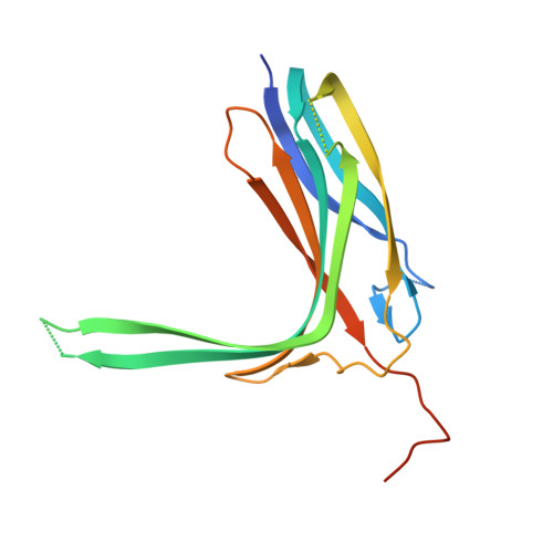
Crystal structure of N-lobe of human ARRDC3(1-180)


Experimental Data Snapshot

  • Method: X-RAY DIFFRACTION
  • Resolution: 2.61 Å
  • R-Value Free: 0.252 
  • R-Value Work: 0.208 
  • R-Value Observed: 0.210 

wwPDB Validation   3D Report Full Report


This is version 1.2 of the entry. See complete history


Literature

Insights into beta 2-adrenergic receptor binding from structures of the N-terminal lobe of ARRDC3.

Qi, S.O'Hayre, M.Gutkind, J.S.Hurley, J.H.

(2014) Protein Sci 23: 1708-1716

  • DOI: https://doi.org/10.1002/pro.2549
  • Primary Citation of Related Structures:  
    4R7V, 4R7X

  • PubMed Abstract: 

    ARRDC3 is one of six known human α-arrestins, and has been implicated in the downregulation of the β2-adrenergic receptor (β2AR). ARRDC3 consists of a two-lobed arrestin fold and a C-terminal tail containing two PPYX motifs. In the current model for receptor downregulation by ARRDC3, the arrestin fold portion is thought to bind the receptor, while the PPXY motifs recruit ubiquitin ligases of the NEDD4 family. Here we report the crystal structures of the N-terminal lobe of human ARRDC3 in two conformations, at 1.73 and 2.8 Å resolution, respectively. The structures reveal a large electropositive region that is capable of binding phosphate ions of crystallization. Residues within the basic patch were shown to be important for binding to β2AR, similar to the situation with β-arrestins. This highlights potential parallels in receptor recognition between α- and β-arrestins.


  • Organizational Affiliation

    Department of Molecular and Cell Biology and California Institute for Quantitative Biosciences, University of California, Berkeley, Berkeley, California, 94720.


Macromolecules
Find similar proteins by:  (by identity cutoff)  |  3D Structure
Entity ID: 1
MoleculeChains Sequence LengthOrganismDetailsImage
Arrestin domain-containing protein 3
A, B
185Homo sapiensMutation(s): 0 
Gene Names: ARRDC3KIAA1376
UniProt & NIH Common Fund Data Resources
Find proteins for Q96B67 (Homo sapiens)
Explore Q96B67 
Go to UniProtKB:  Q96B67
PHAROS:  Q96B67
GTEx:  ENSG00000113369 
Entity Groups  
Sequence Clusters30% Identity50% Identity70% Identity90% Identity95% Identity100% Identity
UniProt GroupQ96B67
Sequence Annotations
Expand
  • Reference Sequence
Small Molecules
Ligands 1 Unique
IDChains Name / Formula / InChI Key2D Diagram3D Interactions
PO4
Query on PO4

Download Ideal Coordinates CCD File 
C [auth A]
D [auth A]
E [auth A]
F [auth A]
G [auth A]
C [auth A],
D [auth A],
E [auth A],
F [auth A],
G [auth A],
H [auth A],
I [auth B],
J [auth B],
K [auth B],
L [auth B],
M [auth B]
PHOSPHATE ION
O4 P
NBIIXXVUZAFLBC-UHFFFAOYSA-K
Experimental Data & Validation

Experimental Data

  • Method: X-RAY DIFFRACTION
  • Resolution: 2.61 Å
  • R-Value Free: 0.252 
  • R-Value Work: 0.208 
  • R-Value Observed: 0.210 
  • Space Group: P 41 21 2
Unit Cell:
Length ( Å )Angle ( ˚ )
a = 107.941α = 90
b = 107.941β = 90
c = 117.778γ = 90
Software Package:
Software NamePurpose
HKL-2000data collection
PHASERphasing
PHENIXrefinement
HKL-2000data reduction
HKL-2000data scaling

Structure Validation

View Full Validation Report


Entry History 

Deposition Data

Revision History  (Full details and data files)

  • Version 1.0: 2014-10-01
    Type: Initial release
  • Version 1.1: 2014-12-10
    Changes: Database references
  • Version 1.2: 2024-02-28
    Changes: Data collection, Database references, Derived calculations