Insights into beta 2-adrenergic receptor binding from structures of the N-terminal lobe of ARRDC3.
Qi, S., O'Hayre, M., Gutkind, J.S., Hurley, J.H.(2014) Protein Sci 23: 1708-1716
- PubMed: 25220262 Search on PubMedSearch on PubMed Central
- DOI: https://doi.org/10.1002/pro.2549
- Primary Citation Related Structures:
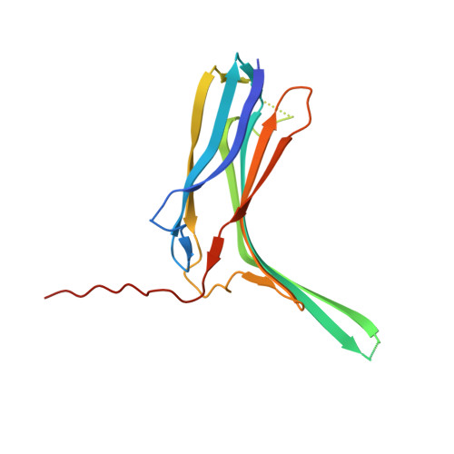
4R7V, 4R7X - PubMed Abstract:
ARRDC3 is one of six known human α-arrestins, and has been implicated in the downregulation of the β2-adrenergic receptor (β2AR). ARRDC3 consists of a two-lobed arrestin fold and a C-terminal tail containing two PPYX motifs. In the current model for receptor downregulation by ARRDC3, the arrestin fold portion is thought to bind the receptor, while the PPXY motifs recruit ubiquitin ligases of the NEDD4 family. Here we report the crystal structures of the N-terminal lobe of human ARRDC3 in two conformations, at 1.73 and 2.8 Å resolution, respectively. The structures reveal a large electropositive region that is capable of binding phosphate ions of crystallization. Residues within the basic patch were shown to be important for binding to β2AR, similar to the situation with β-arrestins. This highlights potential parallels in receptor recognition between α- and β-arrestins.
- Department of Molecular and Cell Biology and California Institute for Quantitative Biosciences, University of California, Berkeley, Berkeley, California, 94720.
Organizational Affiliation: