4R3S

Crystal Structure of anti-MSP2 Fv fragment (mAb6D8)in complex with MSP2 11-23


Experimental Data Snapshot

  • Method: X-RAY DIFFRACTION
  • Resolution: 1.70 Å
  • R-Value Free: 0.200 
  • R-Value Work: 0.152 
  • R-Value Observed: 0.154 

wwPDB Validation   3D Report Full Report


This is version 1.2 of the entry. See complete history


Literature

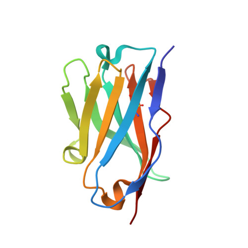
Structural basis for epitope masking and strain specificity of a conserved epitope in an intrinsically disordered malaria vaccine candidate

Morales, R.A.V.MacRaild, C.A.Seow, J.Krishnarjuna, B.Drinkwater, N.Rouet, R.Anders, R.F.Christ, D.McGowan, S.Norton, R.S.

(2015) Sci Rep 5: 10103-10103

  • DOI: https://doi.org/10.1038/srep10103
  • Primary Citation of Related Structures:  
    4QXT, 4QY8, 4QYO, 4R3S

  • PubMed Abstract: 

    Merozoite surface protein 2 (MSP2) is an intrinsically disordered, membrane-anchored antigen of the malaria parasite Plasmodium falciparum. MSP2 can elicit a protective, albeit strain-specific, antibody response in humans. Antibodies are generated to the conserved N- and C-terminal regions but many of these react poorly with the native antigen on the parasite surface. Here we demonstrate that recognition of a conserved N-terminal epitope by mAb 6D8 is incompatible with the membrane-bound conformation of that region, suggesting a mechanism by which native MSP2 escapes antibody recognition. Furthermore, crystal structures and NMR spectroscopy identify transient, strain-specific interactions between the 6D8 antibody and regions of MSP2 beyond the conserved epitope. These interactions account for the differential affinity of 6D8 for the two allelic families of MSP2, even though 6D8 binds to a fully conserved epitope. These results highlight unappreciated mechanisms that may modulate the specificity and efficacy of immune responses towards disordered antigens.


  • Organizational Affiliation

    Medicinal Chemistry, Monash Institute of Pharmaceutical Sciences, Monash University, Parkville, VIC 3052, Australia.


Macromolecules
Find similar proteins by:  (by identity cutoff)  |  3D Structure
Entity ID: 1
MoleculeChains Sequence LengthOrganismDetailsImage
FV FRAGMENT(MAB6D8) HEAVY CHAIN114Mus musculusMutation(s): 0 
Entity Groups  
Sequence Clusters30% Identity50% Identity70% Identity90% Identity95% Identity100% Identity
Sequence Annotations
Expand
  • Reference Sequence
Find similar proteins by:  (by identity cutoff)  |  3D Structure
Entity ID: 2
MoleculeChains Sequence LengthOrganismDetailsImage
FV FRAGMENT(MAB6D8) LIGHT CHAIN111Mus musculusMutation(s): 0 
Entity Groups  
Sequence Clusters30% Identity50% Identity70% Identity90% Identity95% Identity100% Identity
Sequence Annotations
Expand
  • Reference Sequence

Find similar proteins by:  Sequence   |   3D Structure  

Entity ID: 3
MoleculeChains Sequence LengthOrganismDetailsImage
Merozoite surface proteinC [auth Q]15Plasmodium falciparumMutation(s): 2 
UniProt
Find proteins for P50498 (Plasmodium falciparum (isolate 3D7))
Explore P50498 
Go to UniProtKB:  P50498
Entity Groups  
Sequence Clusters30% Identity50% Identity70% Identity90% Identity95% Identity100% Identity
UniProt GroupP50498
Sequence Annotations
Expand
  • Reference Sequence
Experimental Data & Validation

Experimental Data

  • Method: X-RAY DIFFRACTION
  • Resolution: 1.70 Å
  • R-Value Free: 0.200 
  • R-Value Work: 0.152 
  • R-Value Observed: 0.154 
  • Space Group: P 21 21 21
Unit Cell:
Length ( Å )Angle ( ˚ )
a = 35.21α = 90
b = 64.34β = 90
c = 89.87γ = 90
Software Package:
Software NamePurpose
Blu-Icedata collection
PHASERphasing
PHENIXrefinement
MOSFLMdata reduction
SCALAdata scaling

Structure Validation

View Full Validation Report


Entry History 

Deposition Data

Revision History  (Full details and data files)

  • Version 1.0: 2015-06-03
    Type: Initial release
  • Version 1.1: 2019-12-18
    Changes: Advisory, Data collection, Database references, Derived calculations
  • Version 1.2: 2023-11-08
    Changes: Advisory, Data collection, Database references, Refinement description