


Crystal Structure of Dihydroxybenzoate Decarboxylase from Frompolaromonas Sp WITH BOUND MANGANESE AND 2-NITRORESORCINOL
Patskovsky, Y., Vladimirova, A., Toro, R., Bhosle, R., Raushel, F.M., Almo, S.C.To be published.
Experimental Data Snapshot
Starting Model: experimental
View more details
Entity ID: 1 | |||||
|---|---|---|---|---|---|
| Molecule | Chains | Sequence Length | Organism | Details | Image |
| Gamma-resorcylate decarboxylase | 348 | Polaromonas sp. JS666 | Mutation(s): 0 Gene Names: Bpro_2061 EC: 4.1.1.103 | 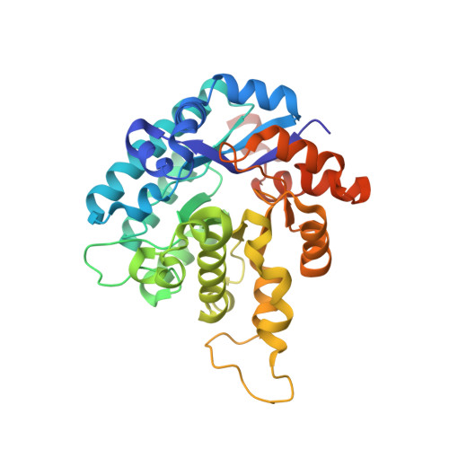 | |
UniProt | |||||
Entity Groups | |||||
| Sequence Clusters | 30% Identity50% Identity70% Identity90% Identity95% Identity100% Identity | ||||
| UniProt Group | Q12BV1 | ||||
Sequence AnnotationsExpand | |||||
Reference Sequence | |||||
| Ligands 5 Unique | |||||
|---|---|---|---|---|---|
| ID | Chains | Name / Formula / InChI Key | 2D Diagram | 3D Interactions | |
| 38L Download:Ideal Coordinates CCD File | CA [auth E] GA [auth F] JA [auth G] M [auth A] PA [auth H] | 2-nitrobenzene-1,3-diol C6 H5 N O4 ZLCPKMIJYMHZMJ-UHFFFAOYSA-N |  | ||
| GOL Download:Ideal Coordinates CCD File | DA [auth F] I [auth A] J [auth A] LA [auth H] N [auth B] | GLYCEROL C3 H8 O3 PEDCQBHIVMGVHV-UHFFFAOYSA-N |  | ||
| BCT Download:Ideal Coordinates CCD File | KA [auth G] | BICARBONATE ION C H O3 BVKZGUZCCUSVTD-UHFFFAOYSA-M |  | ||
| ACT Download:Ideal Coordinates CCD File | AA [auth E] BA [auth E] FA [auth F] IA [auth G] L [auth A] | ACETATE ION C2 H3 O2 QTBSBXVTEAMEQO-UHFFFAOYSA-M |  | ||
| MN Download:Ideal Coordinates CCD File | EA [auth F] HA [auth G] K [auth A] MA [auth H] O [auth B] | MANGANESE (II) ION Mn WAEMQWOKJMHJLA-UHFFFAOYSA-N |  | ||
| Length ( Å ) | Angle ( ˚ ) |
|---|---|
| a = 80.958 | α = 90 |
| b = 151.058 | β = 92.15 |
| c = 143.826 | γ = 90 |
| Software Name | Purpose |
|---|---|
| PHASER | phasing |
| REFMAC | refinement |
| HKL-3000 | data reduction |
| HKL-3000 | data scaling |


