4Q78 | pdb_00004q78

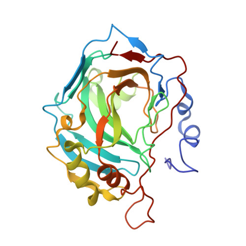
Structure-assisted design of carborane-based inhibitors of carbonic anhydrase


Experimental Data Snapshot

  • Method: X-RAY DIFFRACTION
  • Resolution: 1.00 Å
  • R-Value Free: 
    0.201 (Depositor), 0.206 (DCC) 
  • R-Value Work: 
    0.175 (Depositor), 0.184 (DCC) 
  • R-Value Observed: 
    0.176 (Depositor) 

wwPDB Validation 3D Report Full Report

Validation slider image for 4Q78

Ligand Structure Quality Assessment 


This is version 1.2 of the entry. See complete history. 

Literature

Carborane-based carbonic anhydrase inhibitors: insight into CAII/CAIX specificity from a high-resolution crystal structure, modeling, and quantum chemical calculations.

Mader, P., Pecina, A., Cigler, P., Lepsik, M., Sicha, V., Hobza, P., Gruner, B., Fanfrlik, J., Brynda, J., Rezacova, P.

(2014) Biomed Res Int 2014: 389869-389869

  • DOI: https://doi.org/10.1155/2014/389869
  • Primary Citation Related Structures: 
    4Q78

  • PubMed Abstract: 

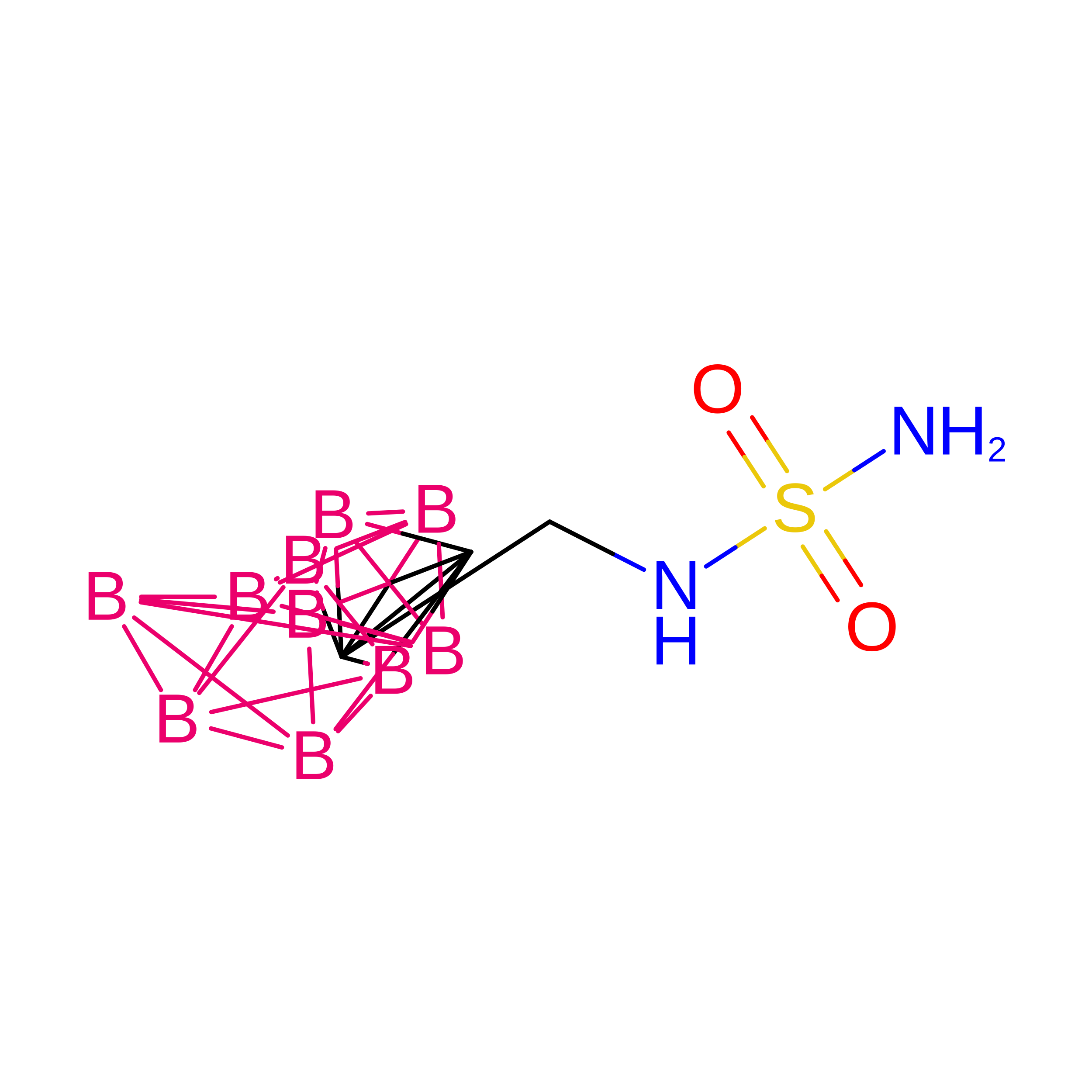
    Carborane-based compounds are promising lead structures for development of inhibitors of carbonic anhydrases (CAs). Here, we report structural and computational analysis applicable to structure-based design of carborane compounds with selectivity toward the cancer-specific CAIX isoenzyme. We determined the crystal structure of CAII in complex with 1-methylenesulfamide-1,2-dicarba-closo-dodecaborane at 1.0 Å resolution and used this structure to model the 1-methylenesulfamide-1,2-dicarba-closo-dodecaborane interactions with CAIX. A virtual glycine scan revealed the contributions of individual residues to the energy of binding of 1-methylenesulfamide-1,2-dicarba-closo-dodecaborane to CAII and CAIX, respectively.


  • Organizational Affiliation: 
    • Institute of Molecular Genetics, Academy of Sciences of the Czech Republic, Vídeňská 1083, 140 00 Prague 4, Czech Republic ; Structural Genomics Consortium, University of Toronto, Toronto, ON, Canada M5G 1L7.

Macromolecule Content 

  • Total Structure Weight: 30.02 kDa 
  • Atom Count: 2,347 
  • Modeled Residue Count: 258 
  • Deposited Residue Count: 258 
  • Unique protein chains: 1

Macromolecules

Find similar proteins by:|  3D Structure
Entity ID: 1
MoleculeChains  Sequence LengthOrganismDetailsImage
Carbonic anhydrase 2258Homo sapiensMutation(s): 0 
EC: 4.2.1.1 (PDB Primary Data), 4.2.1.69 (UniProt)
UniProt & NIH Common Fund Data Resources
Find proteins for P00918 (Homo sapiens)
Explore P00918 
Go to UniProtKB:  P00918
PHAROS:  P00918
GTEx:  ENSG00000104267 
Entity Groups
Sequence Clusters30% Identity50% Identity70% Identity90% Identity95% Identity100% Identity
UniProt GroupP00918
Sequence Annotations
Expand
Reference Sequence

Experimental Data & Validation

Experimental Data

  • Method: X-RAY DIFFRACTION
  • Resolution: 1.00 Å
  • R-Value Free:  0.201 (Depositor), 0.206 (DCC) 
  • R-Value Work:  0.175 (Depositor), 0.184 (DCC) 
  • R-Value Observed: 0.176 (Depositor) 
Space Group: P 1 21 1
Unit Cell:
Length ( Å )Angle ( ˚ )
a = 42.2α = 90
b = 41.725β = 104.36
c = 72.155γ = 90
Software Package:
Software NamePurpose
MOSFLMdata reduction
SCALAdata scaling
REFMACrefinement
PDB_EXTRACTdata extraction

Structure Validation

View Full Validation Report


Ligand Structure Quality Assessment 


Entry History 

Deposition Data

Revision History  (Full details and data files)

  • Version 1.0: 2015-03-18
    Type: Initial release
  • Version 1.1: 2024-02-28
    Changes: Advisory, Data collection, Database references, Derived calculations
  • Version 1.2: 2026-08-12
    Type: Remediation
    Reason: Metalloprotein remediation
    Changes: Derived calculations, Structure summary