4Q43

Polymerase-damaged DNA complex


Experimental Data Snapshot

  • Method: X-RAY DIFFRACTION
  • Resolution: 2.45 Å
  • R-Value Free: 0.268 
  • R-Value Work: 0.214 
  • R-Value Observed: 0.217 

wwPDB Validation   3D Report Full Report


Ligand Structure Quality Assessment 


This is version 1.1 of the entry. See complete history


Literature

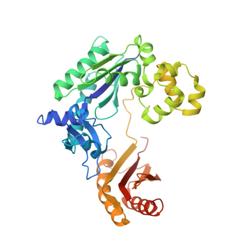
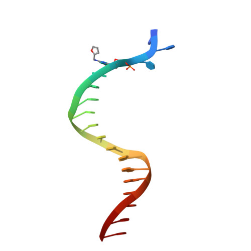
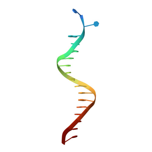
Unique structural features in DNA polymerase IV enable efficient bypass of the N2 adduct induced by the nitrofurazone antibiotic

Kottur, J.Sharma, A.Gore, K.R.Narayanan, N.Samanta, B.Pradeepkumar, P.I.Nair, D.T.

(2015) Structure 23: 56-67

  • DOI: https://doi.org/10.1016/j.str.2014.10.019
  • Primary Citation of Related Structures:  
    4Q43, 4Q44, 4Q45

  • PubMed Abstract: 

    The reduction in the efficacy of therapeutic antibiotics represents a global problem of increasing intensity and concern. Nitrofuran antibiotics act primarily through the formation of covalent adducts at the N(2) atom of the deoxyguanosine nucleotide in genomic DNA. These adducts inhibit replicative DNA polymerases (dPols), leading to the death of the prokaryote. N(2)-furfuryl-deoxyguanosine (fdG) represents a stable structural analog of the nitrofuran-induced adducts. Unlike other known dPols, DNA polymerase IV (PolIV) from E. coli can bypass the fdG adduct accurately with high catalytic efficiency. This property of PolIV is central to its role in reducing the sensitivity of E. coli toward nitrofuran antibiotics such as nitrofurazone (NFZ). We present the mechanism used by PolIV to bypass NFZ-induced adducts and thus improve viability of E. coli in the presence of NFZ. Our results can be used to develop specific inhibitors of PolIV that may potentiate the activity of nitrofuran antibiotics.


  • Organizational Affiliation

    National Centre for Biological Sciences (NCBS-TIFR), GKVK Campus, Bellary Road, Bangalore 560065, India; Manipal University, Manipal.edu, Madhav Nagar, Manipal 576104, India.


Macromolecules

Find similar proteins by:  (by identity cutoff)  |  3D Structure
Entity ID: 1
MoleculeChains Sequence LengthOrganismDetailsImage
DNA polymerase IVA,
B [auth F]
352Escherichia coli K-12Mutation(s): 0 
Gene Names: dinB
EC: 2.7.7.7
UniProt
Find proteins for Q47155 (Escherichia coli (strain K12))
Explore Q47155 
Go to UniProtKB:  Q47155
Entity Groups  
Sequence Clusters30% Identity50% Identity70% Identity90% Identity95% Identity100% Identity
UniProt GroupQ47155
Sequence Annotations
Expand
  • Reference Sequence

Find similar nucleic acids by:  Sequence   |   3D Structure  

Entity ID: 2
MoleculeChains LengthOrganismImage
DNA (5'-D(*TP*CP*TP*(RDG)P*GP*GP*GP*TP*CP*CP*TP*AP*GP*GP*AP*CP*CP*C)-3')C [auth B],
E [auth G]
18synthetic construct
Sequence Annotations
Expand
  • Reference Sequence

Find similar nucleic acids by:  Sequence   |   3D Structure  

Entity ID: 3
MoleculeChains LengthOrganismImage
DNA (5'-D(*T*CP*TP*AP*GP*GP*GP*TP*CP*CP*TP*AP*GP*GP*AP*CP*CP*C)-3')D [auth C],
F [auth H]
18synthetic construct
Sequence Annotations
Expand
  • Reference Sequence
Experimental Data & Validation

Experimental Data

  • Method: X-RAY DIFFRACTION
  • Resolution: 2.45 Å
  • R-Value Free: 0.268 
  • R-Value Work: 0.214 
  • R-Value Observed: 0.217 
  • Space Group: P 1 21 1
Unit Cell:
Length ( Å )Angle ( ˚ )
a = 85.83α = 90
b = 56.79β = 91.96
c = 112.02γ = 90
Software Package:
Software NamePurpose
PHASERphasing
PHENIXrefinement
MOSFLMdata reduction
SCALAdata scaling

Structure Validation

View Full Validation Report


Ligand Structure Quality Assessment 


Entry History 

Deposition Data

Revision History  (Full details and data files)

  • Version 1.0: 2015-05-06
    Type: Initial release
  • Version 1.1: 2023-11-08
    Changes: Data collection, Database references, Derived calculations, Refinement description