Structure of a truncated form of leucine zipper II of JIP3 reveals an unexpected antiparallel coiled-coil arrangement.
Llinas, P., Chenon, M., Nguyen, T.Q., Moreira, C., de Regibus, A., Coquard, A., Ramos, M.J., Guerois, R., Fernandes, P.A., Menetrey, J.(2016) Acta Crystallogr F Struct Biol Commun 72: 198-206
- PubMed: 26919523 Search on PubMedSearch on PubMed Central
- DOI: https://doi.org/10.1107/S2053230X16001576
- Primary Citation Related Structures:
4PXJ - PubMed Abstract:
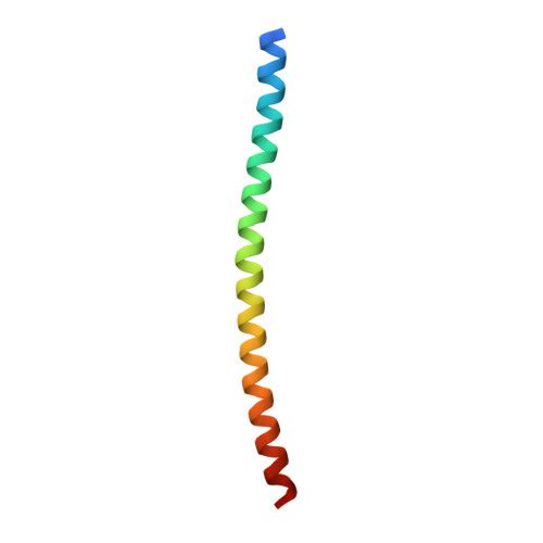
JIP3 and JIP4, two highly related scaffolding proteins for MAP kinases, are binding partners for two molecular motors as well as for the small G protein ARF6. The leucine zipper II (LZII) region of JIP3/4 is the binding site for these three partners. Previously, the crystal structure of ARF6 bound to JIP4 revealed LZII in a parallel coiled-coil arrangement. Here, the crystal structure of an N-terminally truncated form of LZII of JIP3 alone shows an unexpected antiparallel arrangement. Using molecular dynamics and modelling, the stability of this antiparallel LZII arrangement, as well as its specificity for ARF6, were investigated. This study highlights that N-terminal truncation of LZII can change its coiled-coil orientation without affecting its overall stability. Further, a conserved buried asparagine residue was pinpointed as a possible structural determinant for this dramatic structural rearrangement. Thus, LZII of JIP3/4 is a versatile structural motif, modifications of which can impact partner recognition and thus biological function.
- Laboratoire d'Enzymologie et Biochimie Structurales (LEBS), CNRS, Université Paris-Sud, 1 Avenue de la Terrasse, 91190 Gif-sur-Yvette, France.
Organizational Affiliation: