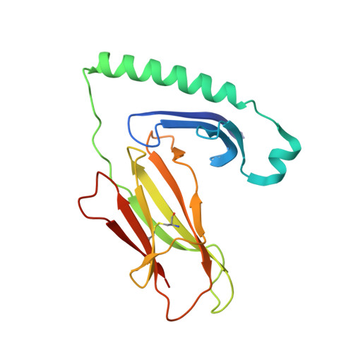
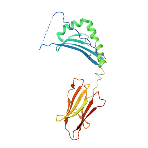
4P57

MHC TCR peptide complex


Experimental Data Snapshot

  • Method: X-RAY DIFFRACTION
  • Resolution: 2.60 Å
  • R-Value Free: 0.245 
  • R-Value Work: 0.208 
  • R-Value Observed: 0.210 

wwPDB Validation   3D Report Full Report


Ligand Structure Quality Assessment 


This is version 1.3 of the entry. See complete history


Literature

Structural basis of chronic beryllium disease: linking allergic hypersensitivity and autoimmunity.

Clayton, G.M.Wang, Y.Crawford, F.Novikov, A.Wimberly, B.T.Kieft, J.S.Falta, M.T.Bowerman, N.A.Marrack, P.Fontenot, A.P.Dai, S.Kappler, J.W.

(2014) Cell 158: 132-142

  • DOI: https://doi.org/10.1016/j.cell.2014.04.048
  • Primary Citation of Related Structures:  
    4P4K, 4P4R, 4P57, 4P5K, 4P5M

  • PubMed Abstract: 

    T-cell-mediated hypersensitivity to metal cations is common in humans. How the T cell antigen receptor (TCR) recognizes these cations bound to a major histocompatibility complex (MHC) protein and self-peptide is unknown. Individuals carrying the MHCII allele, HLA-DP2, are at risk for chronic beryllium disease (CBD), a debilitating inflammatory lung condition caused by the reaction of CD4 T cells to inhaled beryllium. Here, we show that the T cell ligand is created when a Be(2+) cation becomes buried in an HLA-DP2/peptide complex, where it is coordinated by both MHC and peptide acidic amino acids. Surprisingly, the TCR does not interact with the Be(2+) itself, but rather with surface changes induced by the firmly bound Be(2+) and an accompanying Na(+) cation. Thus, CBD, by creating a new antigen by indirectly modifying the structure of preexisting self MHC-peptide complex, lies on the border between allergic hypersensitivity and autoimmunity.


  • Organizational Affiliation

    Howard Hughes Medical Institute, National Jewish Health, Denver, CO 80206, USA; Department of Biomedical Research, National Jewish Health, Denver, CO 80206, USA; Department of Immunology and Microbiology, University of Colorado Denver School of Medicine, Aurora, CO 80045, USA.


Macromolecules
Find similar proteins by:  (by identity cutoff)  |  3D Structure
Entity ID: 1
MoleculeChains Sequence LengthOrganismDetailsImage
HLA class II histocompatibility antigen, DP alpha 1 chain
A, C
183Homo sapiensMutation(s): 0 
Gene Names: HLA-DPA1HLA-DP1AHLASB
UniProt & NIH Common Fund Data Resources
Find proteins for P20036 (Homo sapiens)
Explore P20036 
Go to UniProtKB:  P20036
PHAROS:  P20036
GTEx:  ENSG00000231389 
Entity Groups  
Sequence Clusters30% Identity50% Identity70% Identity90% Identity95% Identity100% Identity
UniProt GroupP20036
Glycosylation
Glycosylation Sites: 2Go to GlyGen: P20036-1
Sequence Annotations
Expand
  • Reference Sequence
Find similar proteins by:  (by identity cutoff)  |  3D Structure
Entity ID: 2
MoleculeChains Sequence LengthOrganismDetailsImage
mim2 peptide,HLA class II histocompatibility antigen, DP beta 1 chain
B, D
212Homo sapiensMutation(s): 2 
UniProt & NIH Common Fund Data Resources
Find proteins for P04440 (Homo sapiens)
Explore P04440 
Go to UniProtKB:  P04440
PHAROS:  P04440
GTEx:  ENSG00000223865 
Entity Groups  
Sequence Clusters30% Identity50% Identity70% Identity90% Identity95% Identity100% Identity
UniProt GroupP04440
Glycosylation
Glycosylation Sites: 1Go to GlyGen: P04440-1
Sequence Annotations
Expand
  • Reference Sequence
Small Molecules
Ligands 3 Unique
IDChains Name / Formula / InChI Key2D Diagram3D Interactions
NAG
Query on NAG

Download Ideal Coordinates CCD File 
E [auth A],
F [auth A],
H [auth B],
J [auth C],
K [auth C]
2-acetamido-2-deoxy-beta-D-glucopyranose
C8 H15 N O6
OVRNDRQMDRJTHS-FMDGEEDCSA-N
GOL
Query on GOL

Download Ideal Coordinates CCD File 
G [auth A]GLYCEROL
C3 H8 O3
PEDCQBHIVMGVHV-UHFFFAOYSA-N
CA
Query on CA

Download Ideal Coordinates CCD File 
I [auth B],
L [auth D]
CALCIUM ION
Ca
BHPQYMZQTOCNFJ-UHFFFAOYSA-N
Experimental Data & Validation

Experimental Data

  • Method: X-RAY DIFFRACTION
  • Resolution: 2.60 Å
  • R-Value Free: 0.245 
  • R-Value Work: 0.208 
  • R-Value Observed: 0.210 
  • Space Group: P 41 21 2
Unit Cell:
Length ( Å )Angle ( ˚ )
a = 102.392α = 90
b = 102.392β = 90
c = 234.33γ = 90
Software Package:
Software NamePurpose
BUSTERrefinement

Structure Validation

View Full Validation Report


Ligand Structure Quality Assessment 


Entry History 

Deposition Data

Revision History  (Full details and data files)

  • Version 1.0: 2014-07-16
    Type: Initial release
  • Version 1.1: 2015-01-07
    Changes: Database references
  • Version 1.2: 2020-07-29
    Type: Remediation
    Reason: Carbohydrate remediation
    Changes: Data collection, Database references, Derived calculations, Other, Refinement description, Source and taxonomy, Structure summary
  • Version 1.3: 2023-12-27
    Changes: Data collection, Database references, Structure summary