


Structure of ECP in complex with citrate ions at 1.50 Angstroms
Blanco, J.A., Boix, E., Moussaoui, M., Salazar, V.A.To be published.
Experimental Data Snapshot
wwPDB Validation 3D Report Full Report

Entity ID: 1 | |||||
|---|---|---|---|---|---|
| Molecule | Chains | Sequence Length | Organism | Details | Image |
| Eosinophil cationic protein | 134 | Homo sapiens | Mutation(s): 1 Gene Names: ECP, RNASE3, RNS3 EC: 3.1.27 | 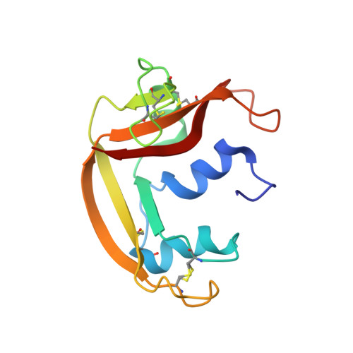 | |
UniProt & NIH Common Fund Data Resources | |||||
PHAROS: P12724 GTEx: ENSG00000169397 | |||||
Entity Groups | |||||
| Sequence Clusters | 30% Identity50% Identity70% Identity90% Identity95% Identity100% Identity | ||||
| UniProt Group | P12724 | ||||
Sequence AnnotationsExpand | |||||
Reference Sequence | |||||
| Ligands 2 Unique | |||||
|---|---|---|---|---|---|
| ID | Chains | Name / Formula / InChI Key | 2D Diagram | 3D Interactions | |
| CIT Download:Ideal Coordinates CCD File | C [auth A], D [auth A], E [auth A], G [auth B], H [auth B] | CITRIC ACID C6 H8 O7 KRKNYBCHXYNGOX-UHFFFAOYSA-N |  | ||
| FE Download:Ideal Coordinates CCD File | F [auth A], I [auth B] | FE (III) ION Fe VTLYFUHAOXGGBS-UHFFFAOYSA-N |  | ||
| Length ( Å ) | Angle ( ˚ ) |
|---|---|
| a = 62.535 | α = 90 |
| b = 62.535 | β = 90 |
| c = 175.05 | γ = 90 |
| Software Name | Purpose |
|---|---|
| PHENIX | refinement |
| Funding Organization | Location | Grant Number |
|---|---|---|
| Ministerio de Economia y Competitividad | Spain | BFU2012-38965 |


