4OWH | pdb_00004owh

PtBr6 binding to HEWL


Experimental Data Snapshot

  • Method: X-RAY DIFFRACTION
  • Resolution: 1.48 Å
  • R-Value Free: 
    0.223 (Depositor), 0.228 (DCC) 
  • R-Value Work: 
    0.190 (Depositor), 0.197 (DCC) 
  • R-Value Observed: 
    0.192 (Depositor) 

wwPDB Validation 3D Report Full Report

Validation slider image for 4OWH

This is version 1.3 of the entry. See complete history

Literature

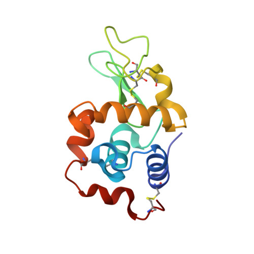
The binding of platinum hexahalides (Cl, Br and I) to hen egg-white lysozyme and the chemical transformation of the PtI6 octahedral complex to a PtI3 moiety bound to His15.

Tanley, S.W.Starkey, L.V.Lamplough, L.Kaenket, S.Helliwell, J.R.

(2014) Acta Crystallogr F Struct Biol Commun 70: 1132-1134

  • DOI: https://doi.org/10.1107/S2053230X14014009
  • Primary Citation Related Structures: 
    4OWC, 4OWE, 4OWH

  • PubMed Abstract: 

    This study examines the binding and chemical stability of the platinum hexahalides K2PtCl6, K2PtBr6 and K2PtI6 when soaked into pre-grown hen egg-white lysozyme (HEWL) crystals as the protein host. Direct comparison of the iodo complex with the chloro and bromo complexes shows that the iodo complex is partly chemically transformed to a square-planar PtI3 complex bound to the N(δ) atom of His15, a chemical behaviour that is not exhibited by the chloro or bromo complexes. Each complex does, however, bind to HEWL in its octahedral form either at one site (PtI6) or at two sites (PtBr6 and PtCl6). As heavy-atom derivatives of a protein, the octahedral shape of the hexahalides could be helpful in cases of difficult-to-interpret electron-density maps as they would be recognisable 'objects'.


  • Organizational Affiliation
    • School of Chemistry, Faculty of Engineering and Physical Sciences, University of Manchester, Brunswick Street, Manchester M13 9PL, England.

Macromolecule Content 

  • Total Structure Weight: 16.3 kDa 
  • Atom Count: 1,152 
  • Modeled Residue Count: 129 
  • Deposited Residue Count: 129 
  • Unique protein chains: 1

Macromolecules

Find similar proteins by:|  3D Structure
Entity ID: 1
MoleculeChains  Sequence LengthOrganismDetailsImage
Lysozyme C129Gallus gallusMutation(s): 0 
EC: 3.2.1.17
UniProt
Find proteins for P00698 (Gallus gallus)
Explore P00698 
Go to UniProtKB:  P00698
Entity Groups
Sequence Clusters30% Identity50% Identity70% Identity90% Identity95% Identity100% Identity
UniProt GroupP00698
Sequence Annotations
Expand
Reference Sequence

Small Molecules

Ligands 5 Unique
IDChains Name / Formula / InChI Key2D Diagram3D Interactions
6BP

Query on 6BP


Download:Ideal Coordinates CCD File
B [auth A],
C [auth A]
HEXABROMOPLATINATE(IV)
Br6 Pt
VOEHEFXBCNCSCK-UHFFFAOYSA-H
BR

Query on BR


Download:Ideal Coordinates CCD File
D [auth A],
E [auth A]
BROMIDE ION
Br
CPELXLSAUQHCOX-UHFFFAOYSA-M
ACT

Query on ACT


Download:Ideal Coordinates CCD File
O [auth A],
P [auth A]
ACETATE ION
C2 H3 O2
QTBSBXVTEAMEQO-UHFFFAOYSA-M
CL

Query on CL


Download:Ideal Coordinates CCD File
F [auth A]
G [auth A]
H [auth A]
I [auth A]
J [auth A]
F [auth A],
G [auth A],
H [auth A],
I [auth A],
J [auth A],
K [auth A],
L [auth A],
M [auth A],
N [auth A]
CHLORIDE ION
Cl
VEXZGXHMUGYJMC-UHFFFAOYSA-M
NA

Query on NA


Download:Ideal Coordinates CCD File
Q [auth A]SODIUM ION
Na
FKNQFGJONOIPTF-UHFFFAOYSA-N

Experimental Data & Validation

Experimental Data

  • Method: X-RAY DIFFRACTION
  • Resolution: 1.48 Å
  • R-Value Free:  0.223 (Depositor), 0.228 (DCC) 
  • R-Value Work:  0.190 (Depositor), 0.197 (DCC) 
  • R-Value Observed: 0.192 (Depositor) 
Space Group: P 43 21 2
Unit Cell:
Length ( Å )Angle ( ˚ )
a = 78.663α = 90
b = 78.663β = 90
c = 36.845γ = 90
Software Package:
Software NamePurpose
REFMACrefinement

Structure Validation

View Full Validation Report


Entry History 

& Funding Information

Deposition Data


Funding OrganizationLocationGrant Number
EPSRCUnited Kingdom--

Revision History  (Full details and data files)

  • Version 1.0: 2014-09-24
    Type: Initial release
  • Version 1.1: 2018-03-07
    Changes: Data collection, Derived calculations, Other, Source and taxonomy, Structure summary
  • Version 1.2: 2023-12-27
    Changes: Data collection, Database references, Refinement description
  • Version 1.3: 2024-11-13
    Changes: Structure summary