4OWA | pdb_00004owa

Carboplatin binding to HEWL under sodium iodide crystallisation conditions


Experimental Data Snapshot

  • Method: X-RAY DIFFRACTION
  • Resolution: 1.80 Å
  • R-Value Free: 
    0.241 (Depositor), 0.243 (DCC) 
  • R-Value Work: 
    0.184 (Depositor), 0.189 (DCC) 
  • R-Value Observed: 
    0.187 (Depositor) 

wwPDB Validation 3D Report Full Report

Validation slider image for 4OWA

This is version 1.2 of the entry. See complete history

Literature

Chemical conversion of cisplatin and carboplatin with histidine in a model protein crystallized under sodium iodide conditions.

Tanley, S.W.Helliwell, J.R.

(2014) Acta Crystallogr F Struct Biol Commun 70: 1127-1131

  • DOI: https://doi.org/10.1107/S2053230X14013995
  • Primary Citation Related Structures: 
    4OWA, 4OWB

  • PubMed Abstract: 

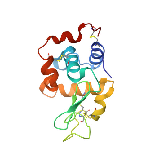
    Cisplatin and carboplatin are platinum anticancer agents that are used to treat a variety of cancers. Previous X-ray crystallographic studies of carboplatin binding to histidine in hen egg-white lysozyme (HEWL) showed a partial chemical conversion of carboplatin to cisplatin owing to the high sodium chloride concentration used in the crystallization conditions. Also, the co-crystallization of HEWL with carboplatin in sodium bromide conditions resulted in the partial conversion of carboplatin to the transbromoplatin form, with a portion of the cyclobutanedicarboxylate (CBDC) moiety still present. The results of the co-crystallization of HEWL with cisplatin or carboplatin in sodium iodide conditions are now reported in order to determine whether the cisplatin and carboplatin converted to the iodo form, and whether this took place in a similar way to the partial conversion of carboplatin to cisplatin in NaCl conditions or to transbromoplatin in NaBr conditions as seen previously. It is reported here that a partial chemical transformation has taken place to a transplatin form for both ligands. The NaI-grown crystals belonged to the monoclinic space group P21 with two molecules in the asymmetric unit. The chemically transformed cisplatin and carboplatin bind to both His15 residues, i.e. in each asymmetric unit. The binding is only at the N(δ) atom of His15. A third platinum species is also seen in both conditions bound in a crevice between symmetry-related molecules. Here, the platinum is bound to three I atoms identified based on their anomalous difference electron densities and their refined occupancies, with the fourth bound atom being a Cl atom (in the cisplatin case) or a portion of the CBDC moiety (in the carboplatin case).


  • Organizational Affiliation
    • School of Chemistry, Faculty of Engineering and Physical Sciences, University of Manchester, Brunswick Street, Manchester M13 9PL, England.

Macromolecule Content 

  • Total Structure Weight: 34.36 kDa 
  • Atom Count: 2,219 
  • Modeled Residue Count: 258 
  • Deposited Residue Count: 258 
  • Unique protein chains: 1

Macromolecules

Find similar proteins by:|  3D Structure
Entity ID: 1
MoleculeChains  Sequence LengthOrganismDetailsImage
Lysozyme C
A, B
129Gallus gallusMutation(s): 0 
EC: 3.2.1.17
UniProt
Find proteins for P00698 (Gallus gallus)
Explore P00698 
Go to UniProtKB:  P00698
Entity Groups
Sequence Clusters30% Identity50% Identity70% Identity90% Identity95% Identity100% Identity
UniProt GroupP00698
Sequence Annotations
Expand
Reference Sequence

Small Molecules

Ligands 4 Unique
IDChains Name / Formula / InChI Key2D Diagram3D Interactions
QPT

Query on QPT


Download:Ideal Coordinates CCD File
NA [auth B],
V [auth A],
W [auth A]
carboplatin
C6 H12 N2 O4 Pt
BHKICZDKIIDMNR-UHFFFAOYSA-L
IOD

Query on IOD


Download:Ideal Coordinates CCD File
AA [auth B]
BA [auth B]
C [auth A]
CA [auth B]
D [auth A]
AA [auth B],
BA [auth B],
C [auth A],
CA [auth B],
D [auth A],
DA [auth B],
E [auth A],
EA [auth B],
F [auth A],
FA [auth B],
G [auth A],
GA [auth B],
H [auth A],
HA [auth B],
I [auth A],
IA [auth B],
J [auth A],
JA [auth B],
K [auth A],
KA [auth B],
L [auth A],
LA [auth B],
M [auth A],
MA [auth B],
N [auth A],
O [auth A],
P [auth A],
Q [auth A],
R [auth A],
S [auth A],
T [auth A],
U [auth A]
IODIDE ION
I
XMBWDFGMSWQBCA-UHFFFAOYSA-M
DMS

Query on DMS


Download:Ideal Coordinates CCD File
OA [auth B]
PA [auth B]
QA [auth B]
X [auth A]
Y [auth A]
OA [auth B],
PA [auth B],
QA [auth B],
X [auth A],
Y [auth A],
Z [auth A]
DIMETHYL SULFOXIDE
C2 H6 O S
IAZDPXIOMUYVGZ-UHFFFAOYSA-N
ACT

Query on ACT


Download:Ideal Coordinates CCD File
RA [auth B]ACETATE ION
C2 H3 O2
QTBSBXVTEAMEQO-UHFFFAOYSA-M

Experimental Data & Validation

Experimental Data

  • Method: X-RAY DIFFRACTION
  • Resolution: 1.80 Å
  • R-Value Free:  0.241 (Depositor), 0.243 (DCC) 
  • R-Value Work:  0.184 (Depositor), 0.189 (DCC) 
  • R-Value Observed: 0.187 (Depositor) 
Space Group: P 1 21 1
Unit Cell:
Length ( Å )Angle ( ˚ )
a = 27.138α = 90
b = 62.561β = 92.59
c = 58.13γ = 90
Software Package:
Software NamePurpose
REFMACrefinement

Structure Validation

View Full Validation Report


Entry History 

& Funding Information

Deposition Data


Funding OrganizationLocationGrant Number
EPSRCUnited Kingdom--

Revision History  (Full details and data files)

  • Version 1.0: 2014-09-24
    Type: Initial release
  • Version 1.1: 2023-12-27
    Changes: Advisory, Data collection, Database references, Derived calculations, Other, Refinement description, Source and taxonomy
  • Version 1.2: 2024-11-20
    Changes: Structure summary