4OQ2

5hmC specific restriction endonuclease PvuRTs1I


Experimental Data Snapshot

  • Method: X-RAY DIFFRACTION
  • Resolution: 2.35 Å
  • R-Value Free: 0.254 
  • R-Value Work: 0.203 
  • R-Value Observed: 0.206 

wwPDB Validation   3D Report Full Report


This is version 1.3 of the entry. See complete history


Literature

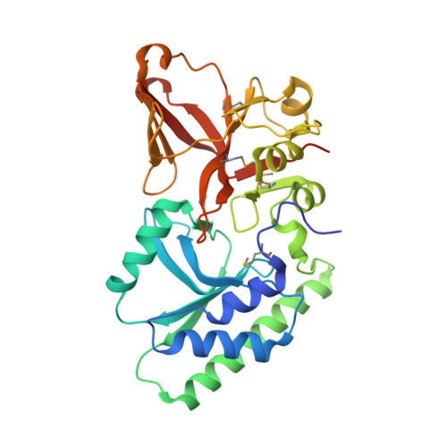
Crystal structure of the 5hmC specific endonuclease PvuRts1I.

Kazrani, A.A.Kowalska, M.Czapinska, H.Bochtler, M.

(2014) Nucleic Acids Res 42: 5929-5936

  • DOI: https://doi.org/10.1093/nar/gku186
  • Primary Citation of Related Structures:  
    4OQ2

  • PubMed Abstract: 

    PvuRts1I is a prototype for a larger family of restriction endonucleases that cleave DNA containing 5-hydroxymethylcytosine (5hmC) or 5-glucosylhydroxymethylcytosine (5ghmC), but not 5-methylcytosine (5mC) or cytosine. Here, we report a crystal structure of the enzyme at 2.35 Å resolution. Although the protein has been crystallized in the absence of DNA, the structure is very informative. It shows that PvuRts1I consists of an N-terminal, atypical PD-(D/E)XK catalytic domain and a C-terminal SRA domain that might accommodate a flipped 5hmC or 5ghmC base. Changes to predicted catalytic residues of the PD-(D/E)XK domain or to the putative pocket for a flipped base abolish catalytic activity. Surprisingly, fluorescence changes indicative of base flipping are not observed when PvuRts1I is added to DNA substrates containing pyrrolocytosine in place of 5hmC (5ghmC). Despite this caveat, the structure suggests a model for PvuRts1I activity and presents opportunities for protein engineering to alter the enzyme properties for biotechnological applications.


  • Organizational Affiliation

    International Institute of Molecular and Cell Biology, Trojdena 4, 02109 Warsaw, Poland mbochtler@iimcb.gov.pl.


Macromolecules
Find similar proteins by:  (by identity cutoff)  |  3D Structure
Entity ID: 1
MoleculeChains Sequence LengthOrganismDetailsImage
Restriction endonuclease PvuRts1 I303Proteus vulgarisMutation(s): 0 
Gene Names: 1239102pvuRts1I
EC: 3.1.21
UniProt
Find proteins for Q52612 (Proteus vulgaris)
Explore Q52612 
Go to UniProtKB:  Q52612
Entity Groups  
Sequence Clusters30% Identity50% Identity70% Identity90% Identity95% Identity100% Identity
UniProt GroupQ52612
Sequence Annotations
Expand
  • Reference Sequence
Small Molecules
Ligands 1 Unique
IDChains Name / Formula / InChI Key2D Diagram3D Interactions
EPE
Query on EPE

Download Ideal Coordinates CCD File 
B [auth A]4-(2-HYDROXYETHYL)-1-PIPERAZINE ETHANESULFONIC ACID
C8 H18 N2 O4 S
JKMHFZQWWAIEOD-UHFFFAOYSA-N
Modified Residues  1 Unique
IDChains TypeFormula2D DiagramParent
MSE
Query on MSE
A
L-PEPTIDE LINKINGC5 H11 N O2 SeMET
Experimental Data & Validation

Experimental Data

  • Method: X-RAY DIFFRACTION
  • Resolution: 2.35 Å
  • R-Value Free: 0.254 
  • R-Value Work: 0.203 
  • R-Value Observed: 0.206 
  • Space Group: P 41 21 2
Unit Cell:
Length ( Å )Angle ( ˚ )
a = 61.99α = 90
b = 61.99β = 90
c = 210.99γ = 90
Software Package:
Software NamePurpose
SHELXDEphasing
DMmodel building
PHENIXmodel building
REFMACrefinement
XDSdata reduction
XSCALEdata scaling
DMphasing
PHENIXphasing

Structure Validation

View Full Validation Report


Entry History 

Deposition Data

Revision History  (Full details and data files)

  • Version 1.0: 2014-03-12
    Type: Initial release
  • Version 1.1: 2014-04-02
    Changes: Database references
  • Version 1.2: 2014-06-04
    Changes: Database references
  • Version 1.3: 2018-03-07
    Changes: Data collection