4OKD

Crystal Structure of Chlamydomonas reinhardtii Isoamylase 1 (ISA1) in complex with maltoheptaose


Experimental Data Snapshot

  • Method: X-RAY DIFFRACTION
  • Resolution: 2.40 Å
  • R-Value Free: 0.227 
  • R-Value Work: 0.192 
  • R-Value Observed: 0.193 

wwPDB Validation   3D Report Full Report


Ligand Structure Quality Assessment 


This is version 2.1 of the entry. See complete history


Literature

Crystal Structure of the Chlamydomonas Starch Debranching Enzyme Isoamylase ISA1 Reveals Insights into the Mechanism of Branch Trimming and Complex Assembly.

Sim, L.Beeren, S.R.Findinier, J.Dauvillee, D.Ball, S.G.Henriksen, A.Palcic, M.M.

(2014) J Biol Chem 289: 22991-23003

  • DOI: https://doi.org/10.1074/jbc.M114.565044
  • Primary Citation of Related Structures:  
    4J7R, 4OKD

  • PubMed Abstract: 

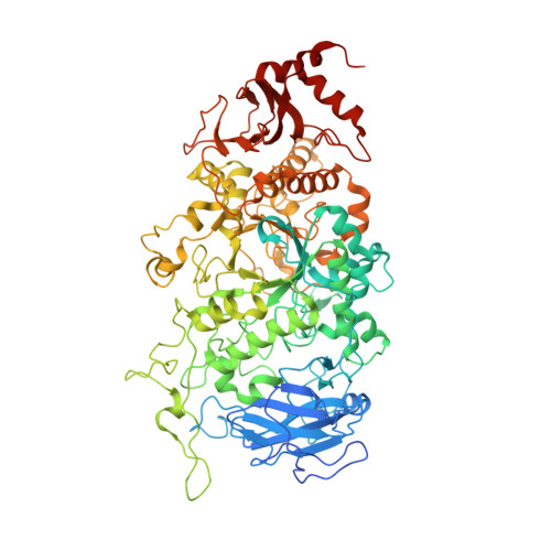
    The starch debranching enzymes isoamylase 1 and 2 (ISA1 and ISA2) are known to exist in a large complex and are involved in the biosynthesis and crystallization of starch. It is suggested that the function of the complex is to remove misplaced branches of growing amylopectin molecules, which would otherwise prevent the association and crystallization of adjacent linear chains. Here, we investigate the function of ISA1 and ISA2 from starch producing alga Chlamydomonas. Through complementation studies, we confirm that the STA8 locus encodes for ISA2 and sta8 mutants lack the ISA1·ISA2 heteromeric complex. However, mutants retain a functional dimeric ISA1 that is able to partly sustain starch synthesis in vivo. To better characterize ISA1, we have overexpressed and purified ISA1 from Chlamydomonas reinhardtii (CrISA1) and solved the crystal structure to 2.3 Å and in complex with maltoheptaose to 2.4 Å. Analysis of the homodimeric CrISA1 structure reveals a unique elongated structure with monomers connected end-to-end. The crystal complex reveals details about the mechanism of branch binding that explains the low activity of CrISA1 toward tightly spaced branches and reveals the presence of additional secondary surface carbohydrate binding sites.


  • Organizational Affiliation

    Carlsberg Laboratory, Gamle Carlsberg Vej 10, DK-1799 Copenhagen V, Denmark and. Electronic address: lya.sim@gmail.com.


Macromolecules
Find similar proteins by:  (by identity cutoff)  |  3D Structure
Entity ID: 1
MoleculeChains Sequence LengthOrganismDetailsImage
Isoamylase
A, B
840Chlamydomonas reinhardtiiMutation(s): 0 
Gene Names: ISA1
EC: 3.2.1.68
UniProt
Find proteins for Q7X8Q2 (Chlamydomonas reinhardtii)
Explore Q7X8Q2 
Go to UniProtKB:  Q7X8Q2
Entity Groups  
Sequence Clusters30% Identity50% Identity70% Identity90% Identity95% Identity100% Identity
UniProt GroupQ7X8Q2
Sequence Annotations
Expand
  • Reference Sequence
Oligosaccharides

Help

Entity ID: 2
MoleculeChains Length2D Diagram Glycosylation3D Interactions
alpha-D-glucopyranose-(1-4)-alpha-D-glucopyranose-(1-4)-alpha-D-glucopyranose-(1-4)-alpha-D-glucopyranose-(1-4)-alpha-D-glucopyranose-(1-4)-alpha-D-glucopyranose-(1-4)-alpha-D-glucopyranose
C, F
7N/A
Glycosylation Resources
GlyTouCan:  G41288IQ
GlyCosmos:  G41288IQ
GlyGen:  G41288IQ
Entity ID: 3
MoleculeChains Length2D Diagram Glycosylation3D Interactions
alpha-D-glucopyranose-(1-4)-alpha-D-glucopyranose-(1-4)-alpha-D-glucopyranose
D, E, G
3N/A
Glycosylation Resources
GlyTouCan:  G96370VA
GlyCosmos:  G96370VA
GlyGen:  G96370VA
Small Molecules
Ligands 1 Unique
IDChains Name / Formula / InChI Key2D Diagram3D Interactions
GLC
Query on GLC

Download Ideal Coordinates CCD File 
H [auth A]alpha-D-glucopyranose
C6 H12 O6
WQZGKKKJIJFFOK-DVKNGEFBSA-N
Biologically Interesting Molecules (External Reference) 1 Unique
Experimental Data & Validation

Experimental Data

  • Method: X-RAY DIFFRACTION
  • Resolution: 2.40 Å
  • R-Value Free: 0.227 
  • R-Value Work: 0.192 
  • R-Value Observed: 0.193 
  • Space Group: P 41 21 2
Unit Cell:
Length ( Å )Angle ( ˚ )
a = 102.75α = 90
b = 102.75β = 90
c = 487.81γ = 90
Software Package:
Software NamePurpose
XSCALEdata scaling
REFMACrefinement
PDB_EXTRACTdata extraction
XDSdata reduction

Structure Validation

View Full Validation Report


Ligand Structure Quality Assessment 


Entry History 

Deposition Data

Revision History  (Full details and data files)

  • Version 1.0: 2014-07-02
    Type: Initial release
  • Version 1.1: 2014-07-23
    Changes: Database references
  • Version 1.2: 2014-09-03
    Changes: Database references
  • Version 2.0: 2020-07-29
    Type: Remediation
    Reason: Carbohydrate remediation
    Changes: Advisory, Atomic model, Data collection, Database references, Derived calculations, Structure summary
  • Version 2.1: 2023-09-20
    Changes: Data collection, Database references, Refinement description, Structure summary